Review
. 2010 Aug;299(2):R416-38.
doi: 10.1152/ajpregu.00111.2010. Epub 2010 May 19.

Neural control of the female urethral and anal rhabdosphincters and pelvic floor muscles

Affiliations
Review

Neural control of the female urethral and anal rhabdosphincters and pelvic floor muscles

Karl B Thor et al. Am J Physiol Regul Integr Comp Physiol. 2010 Aug.

Abstract

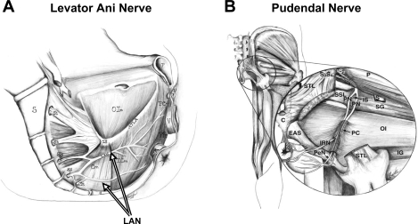
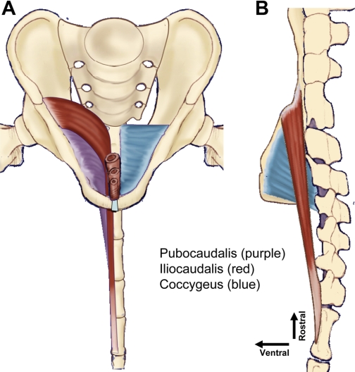
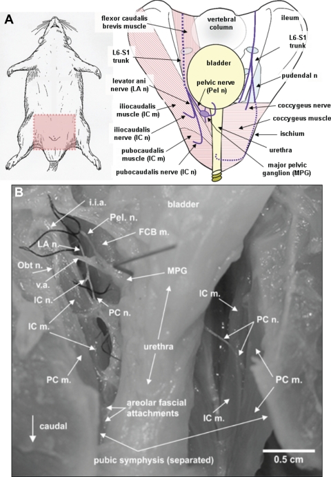
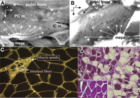
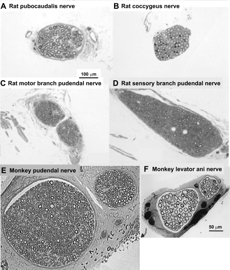
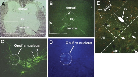
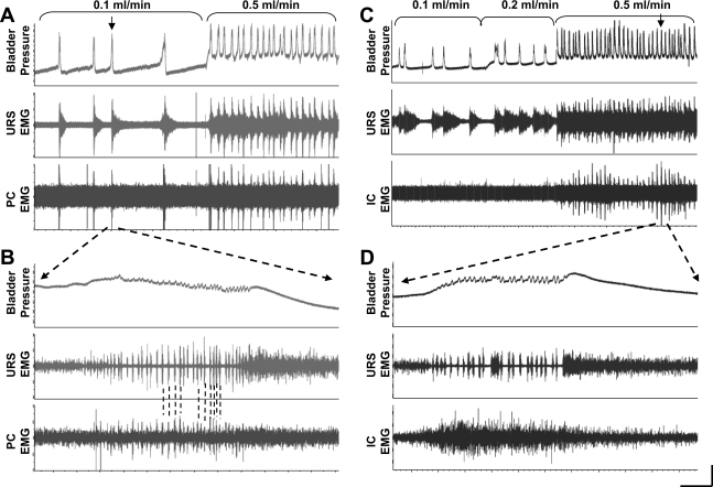
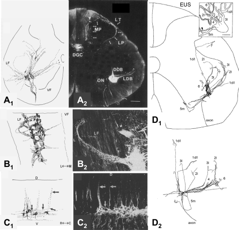
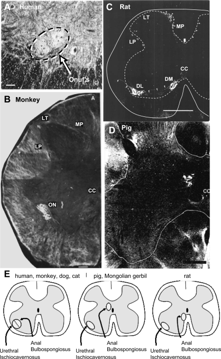
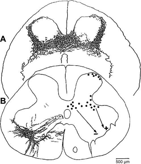
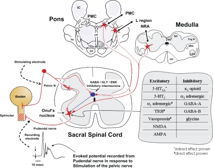
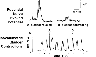
The urethral rhabdosphincter and pelvic floor muscles are important in maintenance of urinary continence and in preventing descent of pelvic organs [i.e., pelvic organ prolapse (POP)]. Despite its clinical importance and complexity, a comprehensive review of neural control of the rhabdosphincter and pelvic floor muscles is lacking. The present review places historical and recent basic science findings on neural control into the context of functional anatomy of the pelvic muscles and their coordination with visceral function and correlates basic science findings with clinical findings when possible. This review briefly describes the striated muscles of the pelvis and then provides details on the peripheral innervation and, in particular, the contributions of the pudendal and levator ani nerves to the function of the various pelvic muscles. The locations and unique phenotypic characteristics of rhabdosphincter motor neurons located in Onuf's nucleus, and levator ani motor neurons located diffusely in the sacral ventral horn, are provided along with the locations and phenotypes of primary afferent neurons that convey sensory information from these muscles. Spinal and supraspinal pathways mediating excitatory and inhibitory inputs to the motor neurons are described; the relative contributions of the nerves to urethral function and their involvement in POP and incontinence are discussed. Finally, a detailed summary of the neurochemical anatomy of Onuf's nucleus and the pharmacological control of the rhabdosphincter are provided.


Figures

Fig. 1.
Fig. 1.
A: sagittal drawing of medial surface of a woman's pelvic floor showing the course of the levator ani nerve (LAN) from the sacral roots (S3–S5) across the internal surface of coccygeus (Cm), iliococcygeus (ICm), puborectalis (PRm), and pubococcygeus (PCm) muscles. S, sacrum; C, coccyx; IS, ischial spine; OIm, obturator internus muscle; ATLA, arcus tendineus levator ani; U, urethra; V, vagina; R, rectum. B: drawing of a posterior view of the hip muscles showing the course of the pudendal nerve (PN) from the S2-S4 roots across the lateral surface of the superior gemellus (SG) and OIm, through the pudendal canal (PC) and its branching into the inferior rectal nerve (IRN) and perineal nerve (PeN). P, periformis muscle; STL, sacroturberous ligament; SSL, sacrospinous ligament; S, sciatic nerve; EAS, external anal sphincter; IG, inferior gemellus muscle. [Adapted from Barber et al. (4).]
Fig. 2.
Fig. 2.
Drawing of origins and insertions of the primary pelvic floor muscles in the rat from the frontal (A) and right side sagital (B) views. In the frontal view (A), the iliocaudalis and the pubocaudalis muscles have been removed from the right side to allow visualization of the underlying coccygeus muscle. The iliocaudalis muscle [origin from the ilium, insertion on caudal vertebrae 5 and 6 (Ca5–6)] is shaded in red, the pubocaudalis muscle (origin from the pubic symphysis; insertion on Ca3–4) is purple, and the coccygeus muscle (origin from the ischium; insertion on Ca1–2) is blue. Note that this arrangement of origins and insertions creates a 3-layered crisscrossed meshing of the muscles from their respective pelvic girdle origins, across each other, to their respective Ca1–6 vertebral insertions.
Fig. 3.
Fig. 3.
A: drawing of rat with pink shaded area (left) as orientation for drawing of female rat pelvis (right). Nerves are shown in purple and their passage behind structures are denoted by dashed lines. The L6-S1 trunk is the origin of the levator ani and pelvic nerves (shown left on the pelvic drawing), which jointly penetrate the pelvic cavity between the flexor caudalis brevis and iliocaudalis muscles. The pelvic nerve then projects ventrally toward the bladder, while the levator ani nerve projects caudally along the plane between the flexor caudalis brevis and the iliocaudalis muscles. The iliocaudalis nerve branches, almost immediately after separation from the pelvic nerve, to penetrate the iliocaudalis muscle, while the pubocaudalis branch continues caudally to penetrate the pubocaudalis muscle. The L6-S1 spinal nerve trunk is also the origin for the pudendal nerve (right on pelvic drawing), which exits the pelvis and travels through the ischiorectal fossa to penetrate the pelvis at the level of the urethral and anal rhabdosphincters. Most of its course is extrapelvic. B: photograph of the pelvis of a female rat showing the relationship between the levator ani nerve and its pubocaudalis and iliocaudalis branches, obturator nerve, pelvic nerve, bladder, and urethra, and the iliocaudalis and pubocaudalis muscles. i.i.a, internal iliac artery; FCB m., flexor caudalis brevis muscle; Obt n., obturator nerve; v.a., vesicular artery; MPG, major pelvic ganglion. [Adapted from Bremer et al. (16).]
Fig. 4.
Fig. 4.
A and B: acetylcholine esterase-stained rat pubocaudalis (PCm), iliocaudalis (ICm), and coccygeus muscle (B) showing a single motor end plate zone (mepz) localized to the midpoint of each muscle. Ct, central tendon of ICm; r, rostral; v, ventral. [Adapted from Bremer et al. (16).] C: levator ani muscle from a squirrel monkey stained with wheat germ agglutinin-rhodamine isothiocyanate showing a muscle spindle and associated intrafusal fibers. (L. M. Pierce and K. B. Thor, unpublished). D: hematoxylin and eosin-stained levator ani muscle taken from a squirrel monkey that had a bilateral levator ani nerve transection 2 yr earlier. Note the shrunken, darkly stained muscle fibers and the fat cell infiltration (unstained areas) compared with the healthy muscle fibers from a control animal (inset, same magnification). Despite significant atrophy of levator ani muscle, this animal did not exhibit POP. [Adapted from Pierce et al. (133).]
Fig. 5.
Fig. 5.
Micrographs of toluidine-stained sections of a rat's pubocaudalis (A) and coccygeus (B) branches of the levator ani nerve; rat's motor (C) and sensory (D) branches of the pudendal nerve; monkey pudendal nerve (E); and monkey levator ani nerve (F). Note the preponderance of large, myelinated motor axons (ca. 10 μm diameter) in the levator ani nerve branches and their absence from pudendal nerve branches. Calibration bar in A applies to A–D, while the calibration bar in F applies to E and F. [A–D adapted from Bremer (16); E–F adapted from Pierce et al. (136).]
Fig. 6.
Fig. 6.
A single transverse sacral section of squirrel monkey spinal cord viewed under brightfield (A) and epifluorescence (B–E) illumination to show levator ani (B and C) and anal rhabdosphincter (D) motor neurons in monkey labeled with retrogradely-transported cholera toxin B (CTB) that had been injected into the levator ani muscle (B and C) or fluorogold that had been injected into the anal rhabdosphincter (D). Note the large (α) and small (γ) CTB-labeled levator ani motor neurons identified by arrows with α and γ labels in C. Also note their CTB-labeled processes distributed in Onuf's nucleus in C. Transganglionically-transported CTB in primary afferent terminals (small green dots in E) in medial lamina VI that overlapped with retrogradely-transported CTB in dendritic bundles of levator ani motor neurons (groups of linear arrays of green dots marked with white arrows), allowing for the possibility of monosynaptic connections. Laminae V, VI, VII, and X are indicated. CC, central canal. The size of the white calibration bar in A represents 200 μm for A and B, 20 μm for C, and 50 μm for D and E. [Adapted from Pierce et al. (135).]
Fig. 7.
Fig. 7.
Examples of pubocaudalis (PC; A and B) and iliocaudalis (IC; C and D) EMG activity during continuous filling cystometry in female rats. Top tracing: bladder pressure; middle tracing: urethral rhabdosphincter (URS) EMG activity; and bottom tracing: PC (A and B) or IC (C and D) EMG activity. The rate of bladder infusion was increased from 0.1 to 0.5 ml/min (A) and 0.1 to 0.2 to 0.5 ml/min (C) as indicated by the brackets. A: note that the URS activity increases preceding the bladder contraction and the activity is maintained for a period of ∼30 s after the contraction, while the PC EMG is only active during the bladder contraction. Increasing the infusion rate produces continuous activity of the URS, while the PC is activated only during the bladder contraction. B: the contraction marked by the arrow in A is at a faster time scale. Note the characteristic phasic bursting pattern of the URS EMG during the high-frequency oscillations (HFOs) of the bladder pressure. Note that the PC EMG also shows phasic activity during the bladder contraction that is temporally correlated with the URS bursting (indicated by dashed lines). C: from a different rat, activity in the IC is not detectable during bladder contractions until the bladder infusion rate is increased to 0.5 ml/min, when the IC EMG is consistently activated with each bladder contraction at the same time as the URS EMG. D: the contraction marked by the arrow in A is at a faster time scale. Note that in contrast to the bursting pattern of the URS during the bladder HFOs that the IC EMG shows only asynchronous, tonic activity with no evidence of bursting. Horizontal calibration bar = 75 s for A and C and 0.5 s for B and D. Vertical calibration bar = 0.5 mV for URS EMG and 0.1 mV for PC and IC EMG, and 15 cm H2O bladder pressure.
Fig. 8.
Fig. 8.
A–C: composite drawings (A1–C1) and photomicrographs (A2–C2) of pudendal motor neurons in cat labeled by application of horseradish peroxidase (HRP) to the pudendal nerve as seen in transverse (A), horizontal (B), and sagittal (C) sections. The photographs provide raw data from 1 of the single sections used to make the corresponding composite drawing. Note that the dendrites of pudendal motor neurons project into the lateral funiculus (LF). D, dorsal; V, ventral; M, medial; L, lateral; R, rostral; C, caudal. Primary afferent terminal labeling can also be seen in Lissauer's tract (LT), the lateral pathway (LP), medial pathway (MP), and dorsal gray commissure (DGC) in A2. (Primary afferents were not drawn in A1, only motor neurons.) DDB, dorsal dendritic bundle; ON, Onuf's nucleus; LDB, lateral dentritic bundle. [Adapted from Thor et al. (171).] D: composite drawing of a single pudendal motor neuron labeled by intracellular injection of HRP showing the transverse (D1) and sagittal (D2) distribution of dendrites (1 d/l, 2l, 3l, 5m). The dashed line in D2 represents the border between the ventral horn and ventral funiculus. Inset in D1 is a 3-D rendition of the neuron. EUS, external urethral sphincter. The arrowhead indicates the cell's axon. [Adapted from Sasaki (148).]
Fig. 9.
Fig. 9.
Interspecies variability in location of anal rhabdosphincter and bulbospongiosus motor neurons in human (A), monkey (B), rat (C), and pig (D). [Adapted from McKenna and Nadelhaft (112), Onufrowicz (128), Roppolo et al. (147), and Blok et al. (9)] C: solid line outlines the spinal cord and dashed line outlines gray matter. E: diagram showing the different distributions of anal and bulbospongiosus motor neurons in various species compared with the similar distribution of urethral and ischiocavernosus motor neurons across species. In D, only the anal rhabdosphincter motor neurons were labeled with retrograde tracer. LT, Lissauer's tract; MP, medial pathway of pudendal afferent fibers; LP, lateral pathway of pudendal afferent fibers; ON, Onuf's nucleus; CC, central canal; DM, dorsomedial nucleus of the pudendal nerve; DL, dorsolateral nucleus of the pudendal nerve. Bars: A = 100 μ; B = 200 μ; C = 500 μ; D = 300 μ.
Fig. 10.
Fig. 10.
A: distribution of afferent projections in the S1 section of the spinal cord from the EUS muscle of the cat. Afferents labeled by anterograde transport of choleratoxin-B-HRP. Composite drawing showing labeled afferent nerves in 5 sequential sections (56 mm thickness) representing an axial distance of 280 mm. B, left: distribution of EUS motor neuron dendrites in the S1 section of cat spinal cord. Neurons were labeled by retrograde transport of choleratoxin-B-HRP. Right: distribution of pseudorabies virus (PRV)-infected interneurons in the S1 section of the spinal cord after injection of PRV into the EUS of the cat. It is likely that lateral interneurons provide an excitatory input (+) and medial interneurons in the dorsal commissure provide an inhibitory input (−) to sphincter motor neurons. [Adapted from de Groat (36).]
Fig. 11.
Fig. 11.
Drawing of proposed model for spinal and supraspinal excitation and inhibition of rhabdosphincter pudendal motor neurons with an example of the evoked potential recorded by an electrode on the pudendal nerve in response to electrical stimulation of the pelvic nerve at 0.5 Hz and a table showing the predominant effects of various receptor subtypes on evoked potentials recorded from the pudendal nerve or urethral rhabdosphincter. Red stellate shapes and lines represent excitatory neurons and their axonal pathways, respectively, while the black oval shape and line represent an inhibitory interneuron and its axonal pathway. Stimulation of the pelvic nerve activates a polysynaptic spinal reflex arc that produces an evoked potential recorded from axons of sphincter motor neurons in Onuf's nucleus at a latency of about 10 ms. In addition, this stimulation also activates inhibitory interneurons that, after 50 ms delay, produce inhibition of sphincter motor neurons for ∼1,000 ms (see Inhibition of Urethral Rhabdosphincter Reflexes During Voiding for details). Presumably this arrangement allows low-frequency pelvic afferent activity (1 Hz) to increase sphincter activity during urine storage and to inhibit sphincter activity when the pelvic afferent activity markedly increases (>5 Hz) as might occur with very large bladder volumes or during a micturition contraction. The model includes GABAergic, glycinergic, or enkephalinergic inhibitory neurons located in the dorsal gray commissure. In addition to spinal excitatory sphincter reflexes, supraspinal pathways originating in the medullary nucleus retroambiguus (NRA) and the pontine L region can activate sphincter motor neurons during Valsalva maneuvers and during urine storage, respectively. When micturition occurs, neurons in the pontine micturition center (PMC) provide descending activation of the GABAergic, glycinergic, or enkephalinergic neurons in the dorsal gray commissure to inhibit sphincter motor neurons and allow voiding to begin. In addition to these predominant pathways, various other areas of the brain (e.g., medullary raphé serotonergic pathways, pontine locus coeruleus noradrenergic pathways, etc.) provide “modulation” of the reflexes. Those excitatory and inhibitory modulatory pathways that have been explored pharmacologically are also listed in the table. For simplicity, the inhibition associated with PMC activation and the inhibition associated with pelvic nerve stimulation are shown passing through the same inhibitory interneuron. However, no evidence yet exists that this is the case. Details and references are in the text and Table 2. Abbreviations: IC, inferior colliculus; NG, nucleus gracilis; NC, nucleus cuneatus; TrigN, spinal nucleus of the trigeminal nerve; LRN, lateral reticular nucleus; pyr, pyramidal tract; mlf, medial longitudinal fasciculus; ll, lateral lemniscus; bc, brachium conjunctivum; bp, brachium pontis; GABA, γ-amino butyric acid; GLY, glycine; ENK, enkephalin; TRH, thyrotropin releasing hormone; NMDA, N-methyl d-aspartate; AMPA, α-amino-3-hydroxy-5-methyl-4-isoxazolepropionic acid; 5-HT, 5-hydroxytryptamine.
Fig. 12.
Fig. 12.
Example of the coordinated inhibition of pudendal reflexes during a bladder contraction. Top: tracings are computer-averaged evoked potentials recorded from the pudendal nerve in response to electrical stimulation (dots) of the pelvic nerve after a latency of 10 ms. Bottom: tracing is recording of bladder pressure under isovolumetric conditions. Note the decreased amplitude of evoked potential B taken during a bladder contraction (at arrow B above bladder pressure tracing) compared with evoked potential A taken between bladder contractions (at arrow A above bladder pressure tracing). [Adapted from Thor et al. (167).]
Fig. 13.
Fig. 13.
Condition-test (C-T; paired-pulse) inhibition of pelvic (PEL) nerve-evoked potentials recorded from URS EMG electrodes. A–C: evoked potentials recorded at interstimulation intervals of 100 ms (left tracings) and 200 ms (right tracings) during control (A) and after the 5-HT1A receptor agonist, 8-OH-DPAT (B) and the 5-HT1A receptor antagonist, WAY100635 (C). Each trace is an average of 10 sweeps from a single animal with drugs administered sequentially. The 2 thin vertical lines on each trace represent the conditioning (first) and test (second) stimuli applied to the pelvic nerve. Note that during the control period at 100 ms, the second stimulus pulse produces virtually no PEL-URS reflex (single arrow at latency for expected evoked potential), while at 200 ms a very small PEL-URS reflex (single arrow) can be seen. Note that 8-OH-DPAT (0.3 mg/kg iv) has modest effects on the amplitude of the conditioning (first) evoked potential but greatly augments the amplitude of the test (second) evoked potential at both 100 and 200 ms interstimulation intervals. Note that WAY100635 (0.3 mg/kg iv) reverses the effect of 8-OH-DPAT. D: graph showing the effects of increasing the C-T interstimulus intervals on the amplitude of the potential evoked by the test (second) stimulus expressed as a percentage of the conditioning (first) stimulus. Recordings were obtained before drugs (control, solid line with squares), after 0.3 mg/kg 8-OH-DPAT (dotted line with triangles), and after WAY100635 (dashed line with circles). *Note that 8-OH-DPAT significantly reduced the inhibition of the conditioning stimulus at the 50 and 100 ms interstimulus intervals, and this effect was completely reversed by WAY100635.
Fig. 14.
Fig. 14.
Example of frequency-response relationship of the URS potentials evoked by pelvic nerve stimulation (top trace). Note the decreased amplitude of the evoked reflex potentials to frequencies ≥ 5 Hz. Bottom trace: 5 Hz stimulation at faster sweep speed. Note the large amplitude reflex evoked by the first stimulus and complete failure of the 2nd–19th evoked responses. Vertical calibration bar = 25 μV. Horizontal calibration bar = 10 s in upper trace and 0.33 s in lower trace.
Fig. 15.
Fig. 15.
Examples of the unique and remarkable association of various neurotransmitters and receptors with pudendal motor neurons in various species. A: cat Leu-enkephalin (Leu-enk) immunoreactivity in an S1 transverse section. Bold arrows point to Onuf's nucleus. DH, dorsal horn. [Adapted from Glazer and Basbaum (55).] B and C: rat 5-HT5A and 5-HT7 receptor immunoreactivity in transverse L6 section. spn, Sacral parasympathetic nucleus; dm, dorsomedial nucleus of the pudendal nerve; dl, dorsolateral of the pudendal nerve; rdl, retrodorsolateral nucleus; vm, ventromedial nucleus. [Adapted from Doly et al. (40) and Doly et al. (41), respectively.] D: neuropeptide Y ([125I]-NPY) autoradiograph of transverse S3 section of human spinal cord. [Adapted from Mantyh et al. (107).] E: growth-associated protein-43 (GAP-43) immunoreactivity in S1 transverse section of human spinal cord. ON, Onuf's nucleus. Open arrow, GAP-43 staining in the dorsal horn. [Adapted from Brook et al. (17).] F: p75 immunoreactivity in a longitudinal L6 section from rat spinal cord through the dorsolateral nucleus of the pudendal nerve. The orientation is similar to that in Fig. 8B except that rostral is toward the right in this panel while 8B1 has rostral to the top. [Adapted from Koliatsos et al. (86).] G: high-power photomicrograph of somatostatin immunoreactivity in Onuf's nucleus (marked by dashed circle) in a transverse S1 section of cat spinal cord. Note that immunoreactivity is restricted to the confines of Onuf's nucleus (K. B. Thor, M. Kawatani, W. C. de Groat, unpublished observation).
Fig. 16.
Fig. 16.
Duloxetine (serotonin norepinephrine reuptake inhibitor) enhances spontaneous sphincter EMG activity and inhibits bladder activity in a chloralose-anesthetized cat. A and B: top tracings are external urethral sphincter (i.e., urethral rhabdosphincter) EMG activity and bottom tracings are bladder pressure recorded during continuous infusion (0.5 ml/min) of 0.5% acetic acid into the bladder. Large downward arrow, infusion begins; small upward arrow (R), release of fluid from urethra. The length of filling from beginning of infusion to release of fluid indicates the bladder capacity. A: control recordings during infusion of dilute acetic acid into the bladder. Note extremely small bladder capacity and very brief bursts of EMG activity after each bladder contraction. B: after administration of 1 mg/kg duloxetine iv, bladder activity is inhibited and bladder capacity increases, while the frequency of sphincter EMG activity is enhanced to the point that individual action potentials are not detectable and appear as a continuous black bar between bladder contractions, while during bladder contractions the EMG activity is virtually abolished. [Adapted from Thor and Katofiasc (168).]
Fig. 17.
Fig. 17.
Ethylketocyclazocine (EKC), a κ-opioid receptor agonist, does not inhibit bladder activity recorded under isovolumetric conditions (A) but inhibits the sphincter reflex recorded from pudendal motor axons in response to electrical stimulation (dots) of afferent axons in the contralateral pudendal nerve (PUD-PUD reflex; B). C: EKC has no effect on the skeletal muscle reflex activated by stimulation of afferent fibers in the sural nerve (SUR) and recorded from motor axons in the nerve to the biceps semimembranosus muscle (BIC). D: effects of high doses of naloxone antagonize EKC on the sphincter reflex. [Adapted from Thor et al. (167).]

Similar articles

Cited by

References

    1. Allen RE, Hosker GL, Smith AR, Warrell DW. Pelvic floor damage and childbirth: a neurophysiological study. Br J Obstet Gynaecol 97: 770–779, 1990 - PubMed
    1. Arvidsson U, Risling M, Frisen J, Piehl F, Fried K, Hokfelt T, Cullheim S. trkC-Like immunoreactivity in the primate descending serotoninergic system. Eur J Neurosci 6: 230–236, 1994 - PubMed
    1. Ashton-Miller JA, DeLancey JO. Functional anatomy of the female pelvic floor. Ann NY Acad Sci 1101: 266–296, 2007 - PubMed
    1. Barber MD, Bremer RE, Thor KB, Dolber PC, Kuehl TJ, Coates KW. Innervation of the female levator ani muscles. Am J Obstet Gynecol 187: 64–71, 2002 - PubMed
    1. Beattie MS, Li Q, Leedy MG, Bresnahan JC. Motoneurons innervating the external anal and urethral sphincters of the female cat have different patterns of dendritic arborization. Neurosci Lett 111: 69–74, 1990 - PubMed

Publication types