. 2010 May 19;2(32):32ra35.
doi: 10.1126/scitranslmed.3001143.

Androgen receptor promotes hepatitis B virus-induced hepatocarcinogenesis through modulation of hepatitis B virus RNA transcription

Affiliations

Androgen receptor promotes hepatitis B virus-induced hepatocarcinogenesis through modulation of hepatitis B virus RNA transcription

Ming-Heng Wu et al. Sci Transl Med. .

Abstract

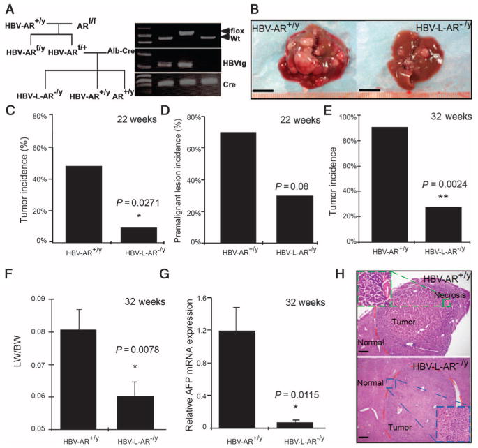
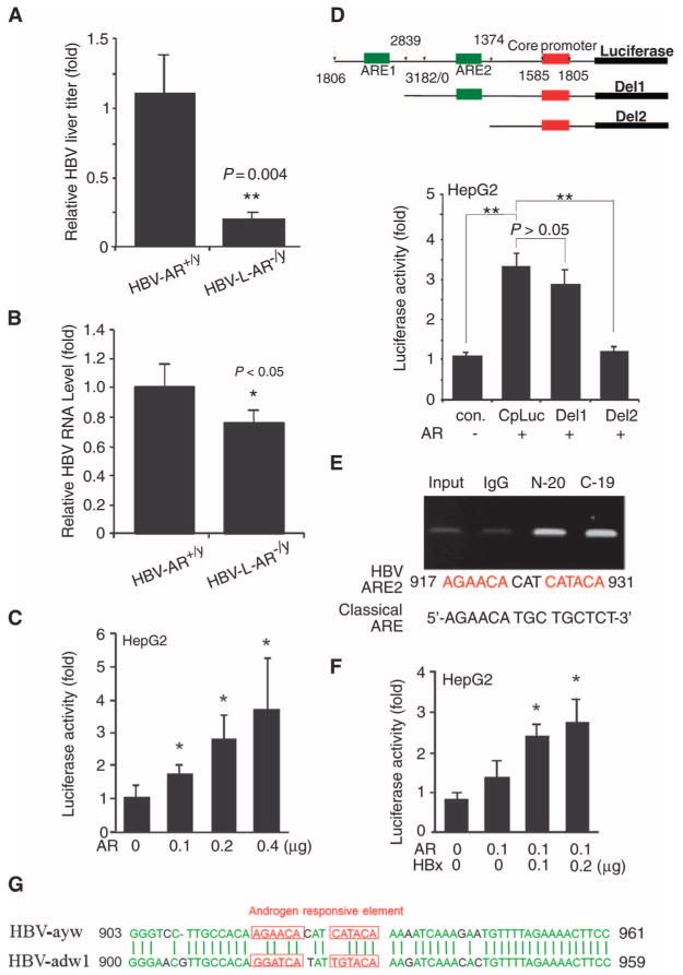
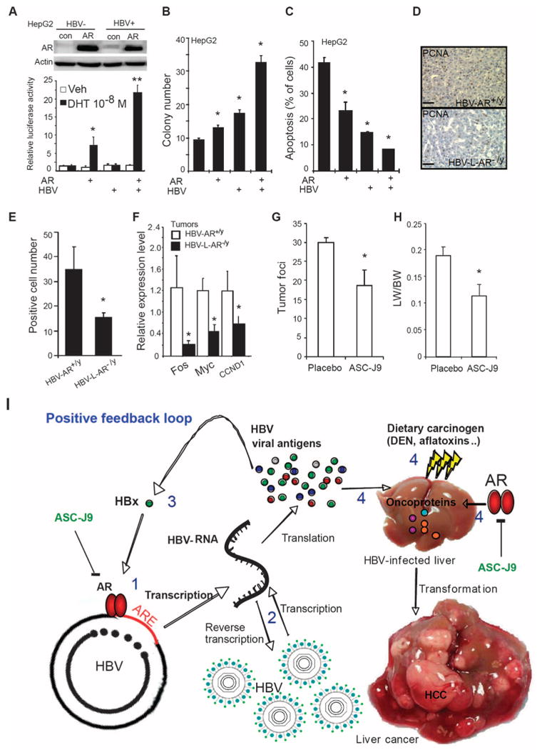
Hepatitis B virus (HBV)-induced hepatitis and carcinogen-induced hepatocellular carcinoma (HCC) are associated with serum androgen concentration. However, how androgen or the androgen receptor (AR) contributes to HBV-induced hepatocarcinogenesis remains unclear. We found that hepatic AR promotes HBV-induced hepatocarcinogenesis in HBV transgenic mice that lack AR only in the liver hepatocytes (HBV-L-AR(-/y)). HBV-L-AR(-/y) mice that received a low dose of the carcinogen N'-N'-diethylnitrosamine (DEN) have a lower incidence of HCC and present with smaller tumor sizes, fewer foci formations, and less alpha-fetoprotein HCC marker than do their wild-type HBV-AR(+/y) littermates. We found that hepatic AR increases the HBV viral titer by enhancing HBV RNA transcription through direct binding to the androgen response element near the viral core promoter. This activity forms a positive feedback mechanism with cooperation with its downstream target gene HBx protein to promote hepatocarcinogenesis. Administration of a chemical compound that selectively degrades AR, ASC-J9, was able to suppress HCC tumor size in DEN-HBV-AR(+/y) mice. These results demonstrate that targeting the AR, rather than the androgen, could be developed as a new therapy to battle HBV-induced HCC.


Conflict of interest statement

Competing interests: ASC-J9 was patented by the University of Rochester, the University of North Carolina, and AndroScience, and then licensed to AndroScience. Both the University of Rochester and C.C. own royalties and equity in AndroScience.

Figures

Fig. 1
Fig. 1
HBV-enhanced hepatocarcinogenesis is suppressed in mice with loss of hepatic AR. (A) Mating scheme of generating AR+/y, HBV-AR+/y, and HBV-L-AR−/y mice. (B) Gross observation of 32-week-old DEN-injected HBV-AR+/y and HBV-L-AR−/y mouse livers. Scale bar, 1.0 cm. (C) Tumor incidence [defined as observable tumors >2 mm in diameter (27)] of 22-week-old DEN-injected HBV-AR+/y (n = 13) and HBV-L-AR−/y (n = 13) mice. (D) Percentage of mice with premalignant liver lesions in 22-week-old DEN-injected HBV-AR+/y (n = 13) and HBV-L-AR−/y (n = 13) mice. (E) Percentage of mice with >20 liver tumor foci in 32-week-old HBV-AR+/y (n = 11) and HBV-L-AR−/y (n = 11) mice injected with DEN. (F) Ratio of liver weight (LW) to whole-body weight (BW) in 32-week-old HBV-L-AR−/y and HBV-AR+/y mice injected with DEN. (G) Liver tumor marker α-AFP mRNA expression in HBV-L-AR−/y mice compared to HBV-AR+/y mice. *P < 0.05, **P < 0.005. Relative α-AFP mRNA expression is normalized by the α-AFP mRNA expression of HBV-AR+/y. (H) Hematoxylin and eosin staining showing representative tumors in the two groups of mice. The tumor from the HBV-AR+/y shows a higher grade with an area of necrosis. Scale bars, 100 μm.
Fig. 2
Fig. 2
AR enhances HBV replication through increase of core promoter activity. (A) HBV DNA was decreased in 22-week-old HBV-L-AR−/y mice (n = 8) compared to HBV-AR+/y mice (n = 11). The relative HBV DNA titer was normalized by the HBV DNA titer of HBV-AR+/y. The P value indicates the comparison between HBV-AR+/y and HBV-L-AR−/y mice. **P < 0.005. (B) HBV mRNA in liver decreased in 32-week-old HBV-L-AR−/y mice as compared to HBV-AR+/y mice. The relative HBV RNA expression was normalized by the HBV RNA of HBV-AR+/y. The P value indicates the comparison between HBV-AR+/y (n = 5) and HBV-L-AR−/y (n = 5) mice. *P < 0.05. (C) AR increases HBV core promoter activity in a dose-dependent manner. *P < 0.05, compared to cells transfected with the vector. Luciferase activities are normalized by Renilla luciferase activity and are shown as mean ± SEM. (D) Top panel: Structure of 5′-deletion constructs of the core promoter. The numbers represent the nucleotide positions in the HBV genome. Green box, putative ARE; red box, HBV pregenome–core promoter. Bottom panel: AR-induced core promoter activity was abolished after deletion of ARE2. Different core promoter constructs were transfected into HepG2 vector or HepG2-AR stable expression cells. Lu-ciferase activity was measured 24 hours after transfection. **P < 0.005. (E) ChIP assay confirms that AR can bind to the ARE2 site of HBV genome. The putative ARE on HBV and classical ARE are compared in the bottom panel. (F) Enhancement of AR-induced core promoter activity by HBx. pSG5, pSG5-AR, core promoter-driven luciferase reporter (CpLuc), pRC/CMV, pCMV-HBx, and pRenilla-TK plasmids were transfected into HepG2 cells in different combinations. Luciferase activities are normalized by Renilla luciferase activity and are shown as mean ± SEM. *P < 0.05, **P < 0.005, compared to cells transfected with vectors. (G) HBV-ayw and HBV-adw1 AREs are of low similarity. Green letters are the aligned conserved sequences, whereas red letters are read as putative ARE on the HBV.
Fig. 3
Fig. 3
AR enhances HBV-promoted hepatocyte transformation. (A) Primary hepatocytes from HBV-AR+/y mice livers have a better passage ability and viability than those from HBV-L-AR−/y mice when cultured in vitro. The hepatocytes were isolated from HBV-AR and HBV-L-AR−/y mice, and cells were continuously propagated until these cells no longer can be passaged. (B) AR stably transfected into BNL CL.2 cells. Mouse testes protein extract served as positive control of AR expression, whereas glyceraldehyde-3-phosphate dehydrogenase (GAPDH) served as loading controls. (C) AR transactivation function was tested by cotransfection of mouse mammary tumor virus (MMTV) luciferase and pRenilla-TK constructs. The luciferase activity was examined after 24 hours of treatment [ethanol (EtOH) or 10 nM dihydrotestosterone (DHT)]. The luciferase activities are normalized by Renilla luciferase activity and are shown as means ± SEM. *P < 0.05, compared with transfected cells treated with ethanol. (D) AR enhances the transformation of HBV-containing hepatocytes. The left top panel shows representative photos of BNL CL.2 cells transformed by SV40 large T antigen, whereas the left bottom panel shows the cells transformed by NMU. The right panel shows the quantitation of colony numbers.
Fig. 4
Fig. 4
AR enhances HBV-mediated tumorigenicity and ASC-J9 suppresses HBV-mediated tumor growth in mice. (A) Top panel: Characterizations of AR expression in HepG2 cells with or without containing the HBV whole genome. AR expression was determined by Western blot. Bottom panel: AR transactivation activity was enhanced by HBV. Cells were cotransfected with pMMTV luciferase reporter and pRenilla-TK plasmid DNA. Luciferase activity was measured 24 hours after treatments (ethanol or 10 nM DHT). *P < 0.05, **P < 0.005, compared with control cells treated with ethanol. (B) AR and HBV cooperatively increased the anchorage-independent growth in HepG2 cells. Colony formation assay was performed in four stable cell lines as described in Materials and Methods. Colonies were stained and counted after 2 weeks of incubation. *P < 0.05, compared with HepG2 control cells. (C) AR and HBV cooperatively decrease stress-induced apoptosis. *P < 0.05, compared with HepG2 control cells. (D) Histological staining of cell proliferation marker PCNA in liver tissues of HBV-AR+/y and HBV-L-AR−/y mice. PCNA signals were measured by immunostaining and representative pictures are shown. Magnification, ×100. Scale bars, 80 μm. (E) Quantitation results of the PCNA-positive staining. Proliferating cells in 32-week-old DEN-injected HBV-AR+/y mice liver tumor are more than HBV-L-AR−/y mice. We counted PCNA-positive cells in three areas (100×) of liver tumor sections of HBV-AR+/y (n = 4) and HBV-L-AR−/y (n = 4) mice. Data are shown as mean ± SEM. The P value indicates the comparison between HBV-AR+/y and HBV-L-AR−/y mice. *P < 0.05. (F) Expression of HCC-associated oncogenes was suppressed in mice with loss of hepatic AR. The 32-week-old mouse liver RNA was extracted and assayed. All the results were shown as mean ± SEM. The P value indicates the comparison between HBV-AR+/y and HBV-L-AR−/y mice. *P < 0.05. (G) ASC-J9 suppresses HBV-induced liver tumor foci in mice. (H) The ratio of liver weight to body weight was reduced in the low-dose, DEN-injected, 36-week-old HBV-AR+/y mice fed with ASC-J9 (50 mg/kg per day) for 45 days. The P value indicates the comparison between placebo and ASC-J9. *P < 0.05. (I) AR promotes HBV-induced hepatocarcinogenesis in a positive feedback manner. The model illustrates that AR induces a positive feed-back response on HBV viral production to mediate hepatocarcinogenesis. (1) Activated AR enhances HBV RNA transcription via binding to ARE on HBV genome. (2) Increased HBV RNA produces more HBV viral antigen and HBV DNA. (3) Increased HBx from AR-enhanced HBV RNA transcription can consequently promote AR-enhanced HBV RNA transcription in a feedback manner. (4) Increased viral antigens in the presence of subminimal dosage of dietary carcinogens can cooperate with AR to promote hepatocyte transformation.

References

    1. Chen CJ, Yu MW, Liaw YF. Epidemiological characteristics and risk factors of hepatocellular carcinoma. J Gastroenterol Hepatol. 1997;12:S294–S308. - PubMed
    1. Chen CJ, Yang HI, Iloeje UH. REVEAL-HBV Study Group, Hepatitis B virus DNA levels and outcomes in chronic hepatitis B. Hepatology. 2009;49:S72–S84. - PubMed
    1. Yu MW, Yang YC, Yang SY, Cheng SW, Liaw YF, Lin SM, Chen CJ. Hormonal markers and hepatitis B virus-related hepatocellular carcinoma risk: A nested case–control study among men. J Natl Cancer Inst. 2001;93:1644–1651. - PubMed
    1. Yu MW, Cheng SW, Lin MW, Yang SY, Liaw YF, Chang HC, Hsiao TJ, Lin SM, Lee SD, Chen PJ, Liu CJ, Chen CJ. Androgen-receptor gene CAG repeats, plasma testosterone levels, and risk of hepatitis B-related hepatocellular carcinoma. J Natl Cancer Inst. 2000;92:2023–2028. - PubMed
    1. Lee CM, Lu SN, Changchien CS, Yeh CT, Hsu TT, Tang JH, Wang JH, Lin DY, Chen CL, Chen WJ. Age, gender, and local geographic variations of viral etiology of hepatocellular carcinoma in a hyperendemic area for hepatitis B virus infection. Cancer. 1999;86:1143–1150. - PubMed

Publication types

MeSH terms