. 2010 Sep;83(3):396-403.
doi: 10.1095/biolreprod.109.083154. Epub 2010 May 19.

Foxa2 is essential for mouse endometrial gland development and fertility

Affiliations

Foxa2 is essential for mouse endometrial gland development and fertility

Jae-Wook Jeong et al. Biol Reprod. 2010 Sep.

Abstract

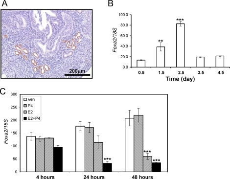
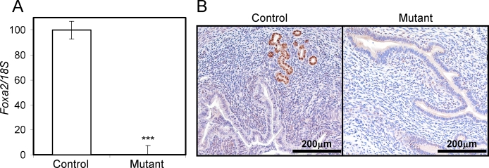
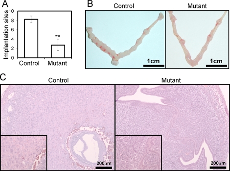
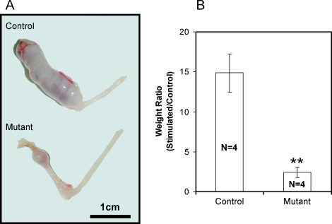
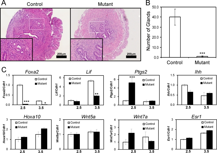
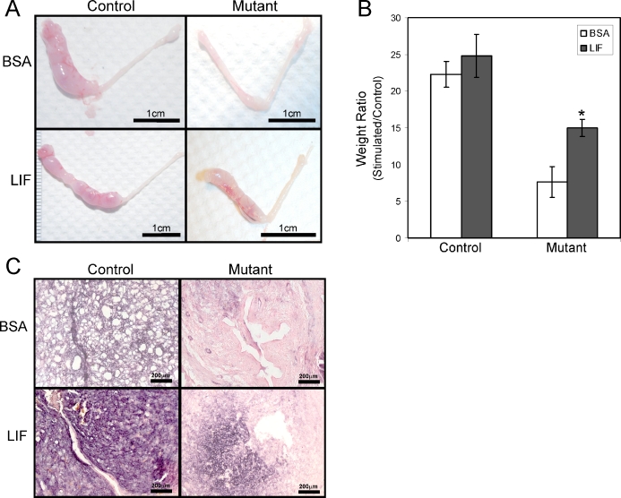
During embryonic development, Foxa2 is required for the formation of the node and notochord, and ablation of this gene results in defects in gastrulation, neural tube patterning, and gut morphogenesis. Foxa2 has been shown to be expressed specifically in the glandular epithelium of the murine uterus. To study the uterine function of Foxa2, this gene was conditionally ablated in the mouse uterus by crossing mice with floxed Foxa2 alleles, Foxa2(loxP/loxP), with the Pgr(cre) mouse model. Pgr(cre/+) Foxa2(loxP/loxP) mice showed significantly reduced fertility. Analysis of the uterus on Day 5.5 of pregnancy showed disrupted blastocyst implantation. Pgr(cre/+) Foxa2(loxP/loxP) mice also showed a severe impairment of the uterus to respond to the artificial induction of the decidual response. Morphological examination of the uteri of these mice showed a severe reduction in the number of endometrial glands. The loss of endometrial glands resulted in the reduction of leukemia inhibitory factor (Lif) expression. The lack of a decidual response could be partially rescued by an intrauterine injection of LIF before the initiation of the decidual response. This analysis demonstrates that Foxa2 regulates endometrial gland development and that mice with a loss of endometrial glands cannot support implantation in part due to the loss of LIF, which is a requisite for fertility in the mouse.


Figures

FIG. 1.
FIG. 1.
The expression pattern of Foxa2 in the murine uterus. A) Localization of FOXA2 in the murine uterus. Eight-week-old female mice were killed. Portions of the uterus were fixed in 4% paraformaldehyde, and immunohistochemistry for FOXA2 was performed. Nuclei are lightly counterstained with hematoxylin. B) The expression pattern of Foxa2 by real-time RT-PCR in pseudopregnancy. Total RNA used for the RT-PCR assays was prepared from the pseudopregnant uteri. The expression level of Foxa2 was measured from Day 0.5 to Day 4.5 in the pseudopregnant uterus. C) The expression pattern of Foxa2 by E2 and P4 in the uterus. Total RNA used for the RT-PCR assays was prepared from wild-type mice that were treated with P4, E2, E2 plus P4, or vehicle (sesame oil) for 4, 24, and 48 h. The results represent the mean ± SEM of three independent RNA sets. **P < 0.01; ***P < 0.001.
FIG. 2.
FIG. 2.
Analysis of Foxa2 conditionally ablated in the murine uterus. The expression level of Foxa2 was measured in the uteri by real-time RT-PCR (A) and immunohistochemistry (B). Eight-week-old control and mutant mice were killed at Day 2.5. Total RNA used for the RT-PCR assays was prepared from the uteri. The results represent the mean ± SEM of three independent RNA sets. ***P < 0.001.
FIG. 3.
FIG. 3.
Implantation defect in the mutant mice. A) Implantation sites in the control and mutant uterus at Day 5.5. Control and mutant mice were killed at Day 5.5. The number of implantation sites was counted in the uteri. The results represent the mean ± SEM of five independent mice. **P < 0.01. B) Gross anatomy of the mutant uteri shows a decrease in the number of implantation sites compared with controls. C) Hematoxylin-eosin staining of control and mutant uteri with blastocysts at Day 5.5. Insets are high-power views of attachment sites.
FIG. 4.
FIG. 4.
Decidualization defect in the mutant mice. Six-week-old mice were ovariectomized and 2 wk later were subjected to a hormone regimen and a decidual stimulus. A) Gross anatomy of the mutant uteri shows a decrease in size of the decidual horn compared with controls. B) Stimulated horn weight:unstimulated horn weight ratio was significantly decreased in the mutant uteri. The results represent the mean ± SEM. **P < 0.01.
FIG. 5.
FIG. 5.
Defect of gland formation in the mutant mice. A) Hematoxylin-eosin staining of uteri from 8-wk-old control and mutant mice. Insets represent high-power views of the boxed region. B) The number of glands was counted from the same area of histological slides. The results represent the mean ± SEM of three independent mice. ***P < 0.001. C) The expression of Lif at Day 2.5 and Day 3.5 in the mutant mice. The expression levels of Foxa2, Lif, Ptgs2, Ihh, Hoxa10, Wnt5a, Wnt7a, and Esr1 were measured at Day 2.5 and Day 3.5 of the pseudopregnant uterus. Total RNA used for the RT-PCR assays was prepared from the pseudopregnant uteri. The results represent the mean ± SEM of three independent RNA sets. *P < 0.05; **P < 0.01; ***P < 0.001.
FIG. 6.
FIG. 6.
Partial rescue of the decidualization defect by recombinant LIF administration in the mutant mice. A) Gross morphology of the decidual response in the mutant uteri 5 days after the decidual stimulus and treatment of 10% BSA or recombinant LIF in 10% BSA. B) Stimulated horn weight:unstimulated horn weight ratio was significantly increased in the mutant uteri treated with recombinant LIF compared with BSA. The results represent the mean ± SEM. *P < 0.05. C) Differentiation by alkaline phosphatase staining in the mutant uterus treated with 10% BSA or recombinant LIF in 10% BSA after the decidual trauma. Bars = 1 cm (A) and 200 μm (C).

Comment in

Similar articles

Cited by

References

    1. Lee KY, DeMayo FJ.Animal models of implantation. Reproduction 2004; 128: 679–695. - PubMed
    1. Gray CA, Bartol FF, Tarleton BJ, Wiley AA, Johnson GA, Bazer FW, Spencer TE.Developmental biology of uterine glands. Biol Reprod 2001; 65: 1311–1323. - PubMed
    1. Gray CA, Taylor KM, Ramsey WS, Hill JR, Bazer FW, Bartol FF, Spencer TE.Endometrial glands are required for preimplantation conceptus elongation and survival. Biol Reprod 2001; 64: 1608–1613. - PubMed
    1. Bell SC, Drife JO.Secretory proteins of the endometrium: potential markers for endometrial dysfunction. Baillieres Clin Obstet Gynaecol 1989; 3: 271–291. - PubMed
    1. Fazleabas AT, Bazer FW, Roberts RM.Purification and properties of a progesterone-induced plasmin/trypsin inhibitor from uterine secretions of pigs and its immunocytochemical localization in the pregnant uterus. J Biol Chem 1982; 257: 6886–6897. - PubMed

Publication types

MeSH terms