. 2010 Jun;120(6):1914-24.
doi: 10.1172/JCI42164. Epub 2010 May 17.

The endothelial cell receptor GRP78 is required for mucormycosis pathogenesis in diabetic mice

Affiliations

The endothelial cell receptor GRP78 is required for mucormycosis pathogenesis in diabetic mice

Mingfu Liu et al. J Clin Invest. 2010 Jun.

Abstract

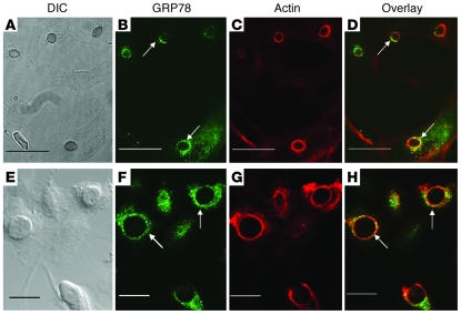
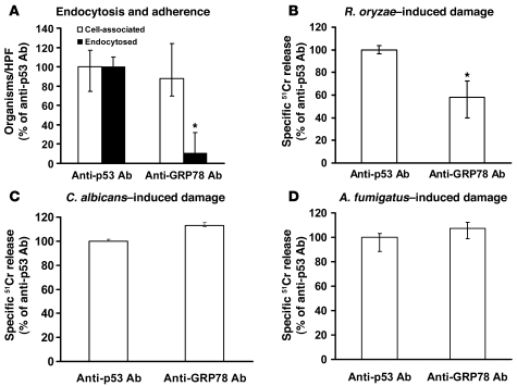
Mucormycosis is a fungal infection of the sinuses, brain, or lungs that causes a mortality rate of at least 50% despite first-line therapy. Because angioinvasion is a hallmark of mucormycosis infections, we sought to define the endothelial cell receptor(s) for fungi of the order Mucorales (the fungi that cause mucormycosis). Furthermore, since patients with elevated available serum iron, including those with diabetic ketoacidosis (DKA), are uniquely susceptible to mucormycosis, we sought to define the role of iron and glucose in regulating the expression of such a receptor. Here, we have identified glucose-regulated protein 78 (GRP78) as what we believe to be a novel host receptor that mediates invasion and damage of human endothelial cells by Rhizopus oryzae, the most common etiologic species of Mucorales, but not Candida albicans or Aspergillus fumigatus. Elevated concentrations of glucose and iron, consistent with those seen during DKA, enhanced GRP78 expression and the resulting R. oryzae invasion and damage of endothelial cells in a receptor-dependent manner. Mice with DKA, which have enhanced susceptibility to mucormycosis, exhibited increased expression of GRP78 in sinus, lungs, and brain compared with normal mice. Finally, GRP78-specific immune serum protected mice with DKA from mucormycosis. These results suggest a unique susceptibility of patients with DKA to mucormycosis and provide a foundation for the development of new therapeutic interventions for these deadly infections.


Figures

Figure 1
Figure 1. Endothelial cell surface GRP78 binds to Mucorales germlings.
(A) Endothelial cell surface proteins were labeled with NHS-biotin (5) and then extracted with n-octyl-β-d-glucopyranoside in PBS containing Ca2+ and Mg2+ and protease inhibitors. The labeled proteins (250 μg) were incubated with germlings (2 × 10⁸) of R. oryzae, then the unbound proteins were removed by extensive rinsing with PBS containing Ca2+ and Mg2+. The membrane proteins that remained bound to the organisms were eluted with 6 M urea, separated on 10% SDS-PAGE, and identified by immunoblotting with an anti-biotin monoclonal Ab. Proteins from another SDS-PAGE were stained with SYPRO Ruby and the bands excised for sequencing. (B) The same membrane that was probed with anti-biotin Ab was stripped and then probed with anti-GRP78 Ab. (C) R. oryzae spores were germinated as previously described (4) for different time intervals, and binding to endothelial cell surface protein and immunoblotting against anti-GRP78 Ab were carried out as in A and B and compared with an equal volume of R. oryzae spores (8 × 10⁸). (D) An immunoblot of endothelial cell surface proteins bound to different Mucorales was developed with an anti-GRP78 Ab. Total membrane, total endothelial cell membrane proteins; M, molecular weight marker.
Figure 2
Figure 2. GRP78 on intact endothelial cells colocalizes with R. oryzae germlings that are being endocytosed.
Confocal microscopic images of endothelial cells infected with R. oryzae cells that have been germinated for 1 hour (AD) or 2 hours (EH). Confluent endothelial cells on a 12-mm-diameter glass coverslip were infected with 105/ml R. oryzae germlings. After 60-minute incubation at 37°C, the cells were fixed with 3% paraformaldehyde, washed, blocked, and then permeabilized (31). The cells were stained with GRP78 using rabbit anti-GRP78 polyclonal Ab (Abcam), followed by a counterstain with goat anti-rabbit IgG conjugated with Alexa Fluor 488 (Molecular Probes, Invitrogen) (B and F). To detect F-actin, the cells were incubated with Alexa Flour 568–labeled phalloidin (Molecular Probes) per the manufacturer’s instructions (C and G). A merged image is shown in D and H. (A and E) The same fields taken with differential interference contrast imaging. Arrows indicate GRP78 and microfilaments that have accumulated around R. oryzae. Scale bars: 30 μm (AD) and 20 μm (EH).
Figure 3
Figure 3. Anti-GRP78 Ab blocks endothelial cell endocytosis of and damage by R. oryzae but not damage caused by C. albicans or A. fumigatus.
Adherence and endocytosis (determined by differential fluorescence) assays were carried out using endothelial cells split on 12-mm glass coverslips, while damage was carried out using the 96-well plate 51Cr release method. Endothelial cells were incubated with 50 μg/ml anti-GRP78 or anti-p53 Ab (control) (Santa Cruz Biotechnology Inc.) for 1 hour prior to addition of R. oryzae germlings. Blocking of GRP78 with Ab abrogates endocytosis of R. oryzae by endothelial cells (data derived from >700 fungal cells interacting with approximately 200 endothelial cells/each group/experiment, with an average of 59% cells being endocytosed in the control) (A) and reduces the ability of the fungus to cause endothelial cell damage (B). However, anti-GRP78 Ab did not block damage caused by C. albicans (C) or A. fumigatus (D). *P < 0.01 compared with anti-p53 Ab by Wilcoxon rank-sum test. n = 6 slides per group from 3 independent experiments for endocytosis, and n = 6 wells per group from 2 independent experiments for damage assay. Data are expressed as median ± interquartile range.
Figure 4
Figure 4. Downregulation of endothelial cell GRP78 expression with shRNA reduces the number of endocytosed organisms and subsequent damage to endothelial cells.
Endothelial cells were transduced with lentivirus containing either shRNA targeting GRP78 or a scrambled sequence (Non-target shRNA). Transduction of endothelial cells with GRP78 shRNA lentiviruses reduced GRP78 transcript levels (A), diminished the number of endocytosed R. oryzae germlings (data derived from >800 fungal cells interacting with approximately 250 endothelial cells/each group/experiment, with an average of 76% being endocytosed in the non-target shRNA) (B), and blocked R. oryzae–induced endothelial cell damage (C). *P < 0.005 compared with non-target shRNA by Wilcoxon rank-sum test for all comparisons. n = 6 slides per group from 3 independent experiments for endocytosis, and n = 6 wells per group from 2 independent experiments for damage assay. Data are expressed as median ± interquartile range.
Figure 5
Figure 5. Heterologous overexpression of GRP78 in CHO cells makes them more susceptible to R. oryzae–induced invasion and subsequent damage.
The C.1 cell line, which was derived from parental DHFR-deficient CHO cells engineered to overexpress GRP78, was found to overexpress GRP78 (A). *P = 0.01 compared with parent cells by nonparametric Wilcoxon rank-sum test; n = 6 per each group. C.1 cells were able to endocytose more R. oryzae germlings (data derived from >950 fungal cells interacting with approximately 300 CHO cells/each group/experiment, with an average of 40.9% being endocytosed in the parent cells) (B) and were more susceptible to R. oryzae–induced damage (C). *P < 0.005 compared with parent cells by Wilcoxon rank-sum test. n = 6 slides per group from 3 independent experiments for endocytosis, and n = 6 wells per group from 2 independent experiments for damage assay. Data are expressed as median ± interquartile range.
Figure 6
Figure 6. Chelation of endothelial cell iron protects the cells from invasion and subsequent damage by R. oryzae.
(A) Endothelial cells were incubated with the iron chelator phenanthroline (60 μM), cytochalasin D (200 nM), or phenanthroline saturated with hemin (20 μM) for 16 hours, then the cells were rinsed and processed for endocytosis and adherence (data derived from >400 fungal cells interacting with approximately 150 endothelial cells/each group/experiment, with an average of 77% being endocytosed in the control). (B) Endothelial cells were treated with varying concentrations of phenanthroline for 16 hours, then the iron chelator was removed prior to carrying out R. oryzae–induced endothelial cell damage. *P < 0.001 versus control (R. oryzae germlings without phenanthroline) by Wilcoxon rank-sum test. n = 6 slides per group from 3 independent experiments for endocytosis, and n = 8 wells per group from 2 independent experiments for damage assay. Data are expressed as median ± interquartile range.
Figure 7
Figure 7. Acidosis as well as iron and glucose concentrations consistent with those seen in DKA patients induce expression of GRP78.
Endothelial cells were incubated at various pHs with or without phenanthroline (A), with iron (B) or glucose (C) concentrations often seen in DKA patients for 5 hours (for studying the effect of acidosis or iron) or 20 hours (for studying the effect of glucose), then the expression of GRP78 was quantified by real-time RT-PCR. n = 6 wells per group from 2 independent experiments. Data are expressed as median ± interquartile range. Cell surface expression of GRP78 on endothelial cells (n = 4 per group from 2 independent experiments) exposed to FeCl3 was quantified using FACS analysis following staining with anti-GRP78 mAb, then counterstaining with anti-mouse Alexa Fluor 488–labeled Ab (D). Data are presented as percent of median fluorescent cells ± interquartile range. *P < 0.01 versus pH 7.4 or the same pH with phenanthroline; **P < 0.05 versus 1 mg/ml glucose or 0 FeCl3 by Wilcoxon rank-sum test.
Figure 8
Figure 8. Iron and glucose concentrations consistent with those seen in DKA patients enhanced invasion of and subsequent damage to endothelial cells by R. oryzae.
Endothelial cells exposed to high concentrations of iron (A) or glucose (B) were subsequently evaluated for their susceptibility to R. oryzae–mediated endocytose and damage. The endocytosis data were derived from more than 600 fungal cells interacting with approximately 200 endothelial cells/each group/experiment, with an average of 51% and 58% endocytosis for no FeCl3 and 1 mg/ml glucose, respectively. *P < 0.01 compared with no FeCl3 or with 1 mg/ml glucose by Wilcoxon rank-sum test. n = 6 slides per group from 3 independent experiments for endocytosis, and n = 9 wells per group from 3 independent experiments for damage assay. Data are expressed as median ± interquartile range.
Figure 9
Figure 9. Anti-GRP78 mAb blocked endothelial cell endocytosis of (A) and subsequent damage by (B) R. oryzae.
Endothelial cells were incubated with R. oryzae in the presence of 50 μg anti-GRP78 Ab or anti-p53 Ab (control). The endocytosis data were derived from more than 500 fungal cells interacting with approximately 150 endothelial cells/each group/experiment, with an average of 71% endocytosis in the control. *P < 0.02 versus anti-p53 Ab. n = 6 slides per group from 3 independent experiments for endocytosis, and n = 6 wells per group from 2 independent experiments for damage assay. Data are expressed as median ± interquartile range.
Figure 10
Figure 10. GRP78 is overexpressed in DKA mice and anti-GRP78 immune serum protects mice from mucormycosis.
(A) Different organs harvested from DKA or normal mice (n = 7 per group) were processed for GRP78 quantification by real-time RT-PCR. *P < 0.05 compared with normal mice. Data are expressed as median ± interquartile range. (B) Survival of mice (n = 18 from 2 independent experiments with similar results) infected intranasally with R. oryzae (105 spores actual inoculum) and treated with anti-GRP78 immune or non-immune sera. **P = 0.037 by log-rank test. The experiment was terminated on day 90, with all remaining mice appearing healthy.

Similar articles

Cited by

References

    1. Ibrahim AS, Edwards JE, Filler SG. Zygomycoses. In: Dismukes WE, Pappas PG, Sobel JD, eds.Clinical Mycology . New York, NY: Oxford University Press; 2003:241–251.
    1. Spellberg B, Edwards J, Jr, Ibrahim A. Novel perspectives on mucormycosis: pathophysiology, presentation, and management. Clin Microbiol Rev. 2005;18(3):556–569. doi: 10.1128/CMR.18.3.556-569.2005. - DOI - PMC - PubMed
    1. Gleissner B, Schilling A, Anagnostopolous I, Siehl I, Thiel E. Improved outcome of zygomycosis in patients with hematological diseases? Leuk Lymphoma. 2004;45(7):1351–1360. doi: 10.1080/10428190310001653691. - DOI - PubMed
    1. Ibrahim AS, Spellberg B, Avanessian V, Fu Y, Edwards JE., Jr Rhizopus oryzae adheres to, is phagocytosed by, and damages endothelial cells in vitro. Infect Immun. 2005;73(2):778–783. doi: 10.1128/IAI.73.2.778-783.2005. - DOI - PMC - PubMed
    1. Isberg RR, Leong JM. Multiple beta 1 chain integrins are receptors for invasin, a protein that promotes bacterial penetration into mammalian cells. Cell. 1990;60(5):861–871. doi: 10.1016/0092-8674(90)90099-Z. - DOI - PubMed

Publication types

MeSH terms