. 2010 Jun;120(6):2040-8.
doi: 10.1172/JCI40253. Epub 2010 May 17.

A retinoic acid-dependent network in the foregut controls formation of the mouse lung primordium

Affiliations

A retinoic acid-dependent network in the foregut controls formation of the mouse lung primordium

Felicia Chen et al. J Clin Invest. 2010 Jun.

Abstract

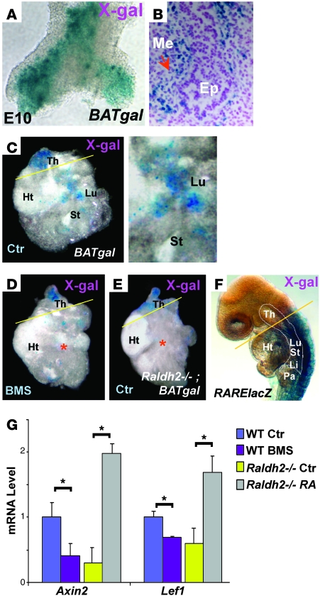
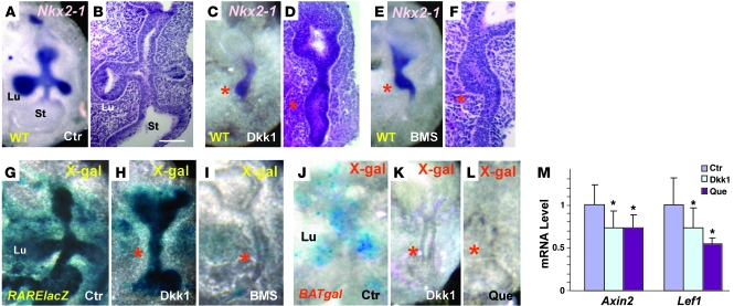
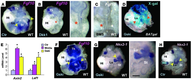
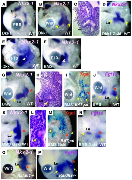
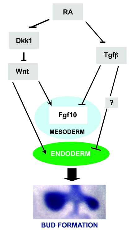
The developmental abnormalities associated with disruption of signaling by retinoic acid (RA), the biologically active form of vitamin A, have been known for decades from studies in animal models and humans. These include defects in the respiratory system, such as lung hypoplasia and agenesis. However, the molecular events controlled by RA that lead to formation of the lung primordium from the primitive foregut remain unclear. Here, we present evidence that endogenous RA acts as a major regulatory signal integrating Wnt and Tgfbeta pathways in the control of Fgf10 expression during induction of the mouse primordial lung. We demonstrated that activation of Wnt signaling required for lung formation was dependent on local repression of its antagonist, Dickkopf homolog 1 (Dkk1), by endogenous RA. Moreover, we showed that simultaneously activating Wnt and repressing Tgfbeta allowed induction of both lung buds in RA-deficient foreguts. The data in this study suggest that disruption of Wnt/Tgfbeta/Fgf10 interactions represents the molecular basis for the classically reported failure to form lung buds in vitamin A deficiency.


Figures

Figure 1
Figure 1. Dkk1 is a target of RA at the onset of lung morphogenesis.
(A) Real-time PCR. Dkk1 upregulation in RA-deficient foreguts (BMS-treated WT and unrescued Raldh2–/–) compared with respective controls (Ctr). (B and C) Dkk1 ISH in WT and Raldh2–/– embryos (12S, top) coimmunostained with Sox2 (bottom), depicting (B) no Dkk1 signals in the foregut (Fg, boxed regions) of WT embryo and (C) strong Dkk1 (blue arrowheads) in both endoderm (red arrowheads, Sox2-labeled brown nuclei) and mesoderm of Raldh2–/– embryo. Original magnification, ×10 (top); ×100 (bottom). (DI) Reciprocal pattern of Dkk1 expression (DF, WMISH) and RARElacZ activity (GI) in the foregut, head, and tail (arrows) in RA-sufficient (D and G, 16S–18S; E and H, 11S–12S), and RA-deficient (F and I, 11S–12S) embryos. (JM) WMISH of Dkk1 in 24-hour cultured foreguts revealed increased signals in RA-deficient conditions in which the lung failed to form (K and M, asterisks). Below, diagrams depict morphology of each condition after 72 hours. Ht, heart; Lu, lung; St, stomach; Th, thyroid. Scale bar: 450 μm (DI); 300 μm (JM).
Figure 2
Figure 2. RA regulation of Wnt during lung formation.
(A and B) E10.0 BATgal lung (A, whole mount; B, section) displaying lacZ activity in both epithelium (Ep) and mesenchyme (Me; red arrow). (CF) Control cultured foregut revealed BATgal activity in the lung and thyroid regions (C) that was suppressed in the lung field of BMS-treated BATgal (D) and Raldh2–/–BATgal (E) foreguts. This activity was preserved in the thyroid region (above yellow lines) of Raldh2–/–BATgal mice (D and E), a domain known to be devoid of RA activity, as seen in RARElacZ reporter mice (F). Li, liver; Pa, pancreas. Original magnification, ×40 (B); ×20 (C, right). (G) Real-time PCR of 24-hour cultured foreguts showing significant downregulation of Axin2 and Lef1 in RA-deficient conditions. *P < 0.05.
Figure 3
Figure 3. Wnt disruption leads to lung bud agenesis in RA-sufficient foreguts.
(AF) Nkx2-1 WMISH (A, C, and E) and corresponding H&E (B, D, and F) of cultured foreguts. Control foreguts showed 2 emerging lung buds labeled by Nkx2-1 (A and B). In Dkk1-treated WT foreguts, Nkx2-1 still marked the lung field, but no buds were present (C and D), reminiscent of BMS-treated WT foreguts (E and F). (GI) X-gal staining demonstrated strong RARElacZ signals in both control and Dkk1-treated foreguts (G and H), in contrast to the lack of signals in the BMS-treated foreguts (I). (JL) BATgal activity was abolished with Dkk1 (K) or quercetin (Que; L) treatment compared with the control (J). Red asterisks in CF, H, I, K, and L mark the prospective lung field. Original magnification, ×10 (A, C, E, and GL). Scale bar: 250 μm (B, D, and F). (M) Real-time PCR revealed downregulation of Axin2 and Lef1 in 24-hour cultured foreguts treated with Dkk1 or quercetin. *P < 0.05 versus control.
Figure 4
Figure 4. Wnt regulates Fgf10 in the foregut mesoderm.
(AC) WMISH of Fgf10 showed local expression in the mesoderm associated with lung buds (A, arrowheads). Dkk1 inhibition of Wnt signaling abolished Fgf10 in the prospective lung field (B, asterisk; enlarged at right), an effect strikingly similar to that of BMS treatment in the foregut (C, asterisk). (D) Hyperactivation of Wnt by Gsk3b inhibitor (Gski) resulted in widespread LacZ activity in BATgal foreguts (red arrowheads). (E) Real-time PCR revealed upregulation of Axin2 and Lef1 in 24-hour cultured foregut treated with Gsk3b inhibitor or Wnt3a. *P < 0.05 versus control. (F) WMISH of Gsk3b inhibitor–treated foregut exhibiting stronger and broader Fgf10 signals (yellow arrowheads) compared with the control (A). (G and H) WMISH of Nkx2-1 in Gsk3b inhibitor–treated (G) or Wnt3a bead–engrafted (H) WT foreguts demonstrated ectopic buds in the lung and stomach (yellow arrowheads). Scale bar: 300 μm (B, right, C, and G).
Figure 5
Figure 5. Wnt rescues lung agenesis in Dkk1-treated and RA-deficient foreguts.
(AD) Engraftment of beads soaked in Wnt3a, but not PBS, rescued lung buds bilaterally in Dkk1-treated foreguts (AC), as confirmed by strong Nkx2-1 expression (arrowheads). Bilateral rescue was also observed when Gsk3b inhibitor was added to Dkk1-treated foreguts (D). (E and F) Exogenous FGF10 (F10) beads induced buds expressing Nkx2-1 (arrows) in Dkk1-treated foreguts (E), a response similarly elicited in BMS-treated foreguts (F). (GP) Engraftment of Wnt3a beads onto BMS-treated foreguts induced only 1 lung bud, labeled by Nkx2-1 (G and H, arrowheads), although BATgal activity (I) and Fgf10 expression (J) were clearly present on both sides of the lung endoderm (arrows). Similar phenotypes were seen in foreguts treated simultaneously with BMS and Gsk3b inhibitor (KN) and in Raldh2–/– foreguts treated with Wnt3a beads (OP). Red asterisks indicate sites of unilateral bud agenesis. Original magnification, ×10 (A, B, DG, IK, and MP); ×20 (C, H, and L).
Figure 6
Figure 6. Tgfβ inhibition and Wnt activation fully rescue lung buds in RA-deficient conditions.
(AH) Addition of Tgfβ blocking antibody (TAb) and Wnt3a bead to BMS-treated foreguts elicited bilateral lung bud formation (A and B, arrowheads), which was associated with local rescue of Fgf10 (C and D, arrowheads and circled regions). Similar rescue of local budding (EG) and Fgf10 expression (H) was observed in Raldh2–/– foreguts treated with the Tgfβ receptor 1 antagonist SB4 and Gsk3b inhibitor. Corresponding H&E staining is shown in B and G. Original magnification, ×20 (A, right, B, and F); ×10 (D and H). Scale bar: 250 μm.
Figure 7
Figure 7. An RA-dependent network in the foregut controls formation of the lung primordium.
During primary lung bud morphogenesis, RA signaling in the foregut mesoderm allows induction of the Wnt pathway by suppressing Dkk1 expression; RA also inhibits Tgfβ signaling. The balanced activity of Wnt and Tgfβ leads to proper mesodermal Fgf10 expression required for formation of the lung primordium. In the foregut endoderm, the RA regulation of Dkk1, and ultimately Wnt, contributes to maintenance of lung progenitor cell fate. RA may also influence the response of the foregut endoderm to Fgf10, presumably through a Tgfβ target asymmetrically distributed in the left-right aspect of endoderm.

Similar articles

Cited by

References

    1. Naik-Mathuria B, Olutoye OO. Foregut abnormalities. Surg Clin North Am. 2006;86(2):261–284, viii. doi: 10.1016/j.suc.2005.12.011. - DOI - PubMed
    1. Gavrilova R, et al. Vitamin A deficiency in an infant with PAGOD syndrome. Am J Med Genet A. 2009;149A(10):2241–2247. - PubMed
    1. Golzio C, et al. Matthew-Wood syndrome is caused by truncating mutations in the retinol-binding protein receptor gene STRA6. Am J Hum Genet. 2007;80(6):1179–1187. doi: 10.1086/518177. - DOI - PMC - PubMed
    1. Wilson JG, Roth CB, Warkany J. An analysis of the syndrome of malformations induced by maternal Vitamin A deficiency. Effects of restoration of Vitamin A at various times during gestation. Am J Anat. 1953;92(2):189–217. doi: 10.1002/aja.1000920202. - DOI - PubMed
    1. Kastner P, et al. Genetic evidence that the retinoid signaling is transduced by heterodimeric RXR/RAR functional units during mouse development. Development. 1997;124(2):313–326. - PubMed

Publication types

MeSH terms