

Deficiency in the serum-derived hyaluronan-associated protein-hyaluronan complex enhances airway hyperresponsiveness in a murine model of asthma
- PMID: 20484920
- PMCID: PMC2945275
- DOI: 10.1159/000314362
Deficiency in the serum-derived hyaluronan-associated protein-hyaluronan complex enhances airway hyperresponsiveness in a murine model of asthma
Abstract
Background: Serum-derived hyaluronan (HA)-associated proteins (SHAPs), the heavy chains of inter-α-trypsin inhibitor, covalently bind to HA to form the SHAP-HA complex. The SHAP-HA complex is involved in the pathophysiology of inflammatory diseases, including rheumatoid arthritis. We investigated whether this complex is also involved in airway allergy.
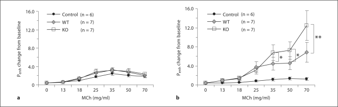
Methods: SHAP-HA-deficient (bikunin knockout, KO) mice and wild-type (WT) mice were immunized twice by intraperitoneal injection of ovalbumin (OVA) and exposed to aerosol OVA for 30 min each day for 2 weeks. Twenty-four hours after the final OVA challenge, airway responsiveness to inhaled methacholine (MCh) was measured, and analysis of bronchoalveolar lavage fluid (BALF) and lung histological studies were done.
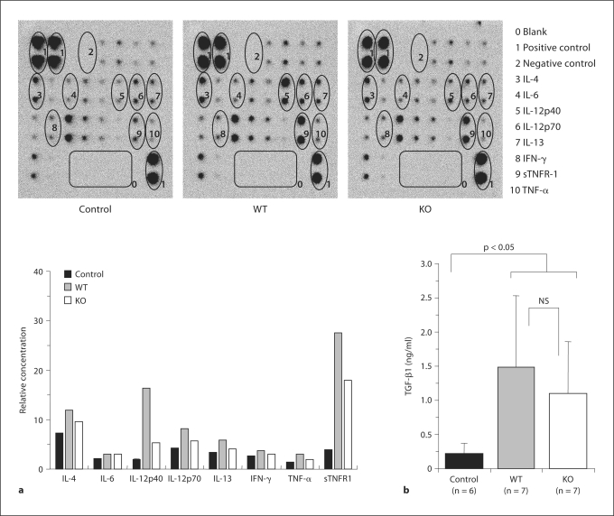
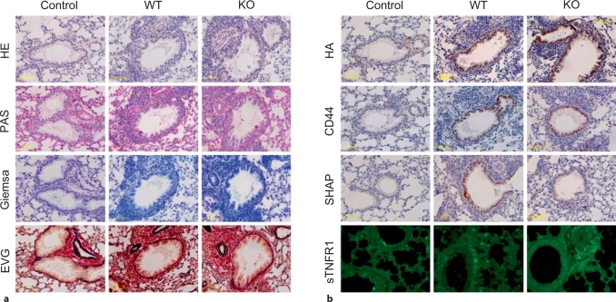
Results: Compared to WT mice, KO mice showed higher airway hyperresponsiveness to inhaled MCh and higher late-phase responses to OVA whereas the early-phase responses were similar. Cell differentials of BALF showed an increased number of macrophages and neutrophils in KO mice. Furthermore, decreased concentrations of soluble tumor necrosis factor receptor-1 (sTNFR1) were found in BALF from KO mice whereas the levels of Th1 and Th2 cytokines were not different from WT mice. Immunochemical study of the lung tissues revealed stronger staining of sTNFR1 in KO than in WT mice.
Conclusions: Our results suggest that in this murine asthma model, the SHAP-HA complex has an inhibitory role in the development of airway hyperresponsiveness and allergic airway inflammation which may be attributed, at least in part, to negative feedback mechanisms exerted by sTNFR1, the shedding of which from the cell surface might also be promoted by the SHAP-HA complex.
Copyright © 2010 S. Karger AG, Basel.
Figures


Similar articles
-
Annexin-1-deficient mice exhibit spontaneous airway hyperresponsiveness and exacerbated allergen-specific antibody responses in a mouse model of asthma.Clin Exp Allergy. 2011 Dec;41(12):1793-803. doi: 10.1111/j.1365-2222.2011.03855.x. Epub 2011 Sep 20. Clin Exp Allergy. 2011. PMID: 22092555
-
Role of thrombin-activatable fibrinolysis inhibitor in allergic bronchial asthma.Lung. 2012 Apr;190(2):189-98. doi: 10.1007/s00408-011-9337-9. Epub 2011 Oct 25. Lung. 2012. PMID: 22037793
-
Petiveria alliacea Suppresses Airway Inflammation and Allergen-Specific Th2 Responses in Ovalbumin-Sensitized Murine Model of Asthma.Chin J Integr Med. 2018 Dec;24(12):912-919. doi: 10.1007/s11655-018-2566-5. Epub 2018 Oct 19. Chin J Integr Med. 2018. PMID: 30341485
-
L-Selectin is required for the development of airway hyperresponsiveness but not airway inflammation in a murine model of asthma.J Allergy Clin Immunol. 2001 Jun;107(6):1019-24. doi: 10.1067/mai.2001.114703. J Allergy Clin Immunol. 2001. PMID: 11398079
-
Comparison of asthma phenotypes in OVA-induced mice challenged via inhaled and intranasal routes.BMC Pulm Med. 2019 Dec 10;19(1):241. doi: 10.1186/s12890-019-1001-9. BMC Pulm Med. 2019. PMID: 31823765 Free PMC article.
Cited by
-
Defining the versican interactome in lung health and disease.Am J Physiol Cell Physiol. 2022 Aug 1;323(2):C249-C276. doi: 10.1152/ajpcell.00162.2022. Epub 2022 Jun 1. Am J Physiol Cell Physiol. 2022. PMID: 35649251 Free PMC article. Review.
-
Urine inter-alpha-trypsin inhibitor family-related proteins may serve as biomarkers for disease activity of lupus.J Clin Lab Anal. 2022 Sep;36(9):e24622. doi: 10.1002/jcla.24622. Epub 2022 Jul 23. J Clin Lab Anal. 2022. PMID: 35870194 Free PMC article.
-
The Hyaluronic Acid-HDAC3-miRNA Network in Allergic Inflammation.Front Immunol. 2015 Apr 30;6:210. doi: 10.3389/fimmu.2015.00210. eCollection 2015. Front Immunol. 2015. PMID: 25983734 Free PMC article. Review.
-
Inhibition of hyaluronan retention by 4-methylumbelliferone suppresses osteosarcoma cells in vitro and lung metastasis in vivo.Br J Cancer. 2011 Dec 6;105(12):1839-49. doi: 10.1038/bjc.2011.459. Epub 2011 Nov 1. Br J Cancer. 2011. PMID: 22045192 Free PMC article.
-
The role of hyaluronan in the pathobiology and treatment of respiratory disease.Am J Physiol Lung Cell Mol Physiol. 2016 May 1;310(9):L785-95. doi: 10.1152/ajplung.00168.2015. Epub 2016 Jan 8. Am J Physiol Lung Cell Mol Physiol. 2016. PMID: 26747781 Free PMC article. Review.
References
-
- Fahy JV, Corry DB, Boushey HA. Airway inflammation and remodeling in asthma. Curr Opin Pulm Med. 2000;6:15–20. - PubMed
-
- Jeffery PK. Remodeling in asthma and chronic obstructive lung disease. Am J Respir Crit Care Med. 2001;164:S28–S38. - PubMed
-
- Umetsu DT, McIntire JJ, Akbari O, Macaubas C, DeKruyff RH. Asthma: An epidemic of dysregulated immunity. Nat Immunol. 2002;3:715–720. - PubMed
-
- Hirst SJ, Twort CH, Lee TH. Differential effects of extracellular matrix proteins on human airway smooth muscle cell proliferation and phenotype. Am J Respir Cell Mol Biol. 2000;23:335–344. - PubMed
Publication types
MeSH terms
Substances
Grants and funding
LinkOut - more resources
Full Text Sources
Other Literature Sources
Medical
Research Materials
