. 2010 Jun 17;465(7300):885-90.
doi: 10.1038/nature09121. Epub 2010 May 19.

Helical assembly in the MyD88-IRAK4-IRAK2 complex in TLR/IL-1R signalling

Affiliations

Helical assembly in the MyD88-IRAK4-IRAK2 complex in TLR/IL-1R signalling

Su-Chang Lin et al. Nature. .

Abstract

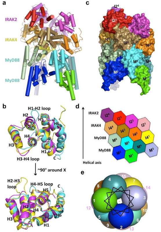
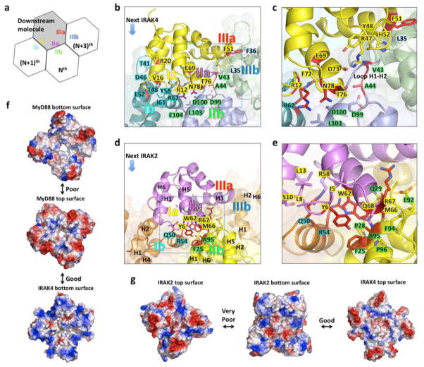
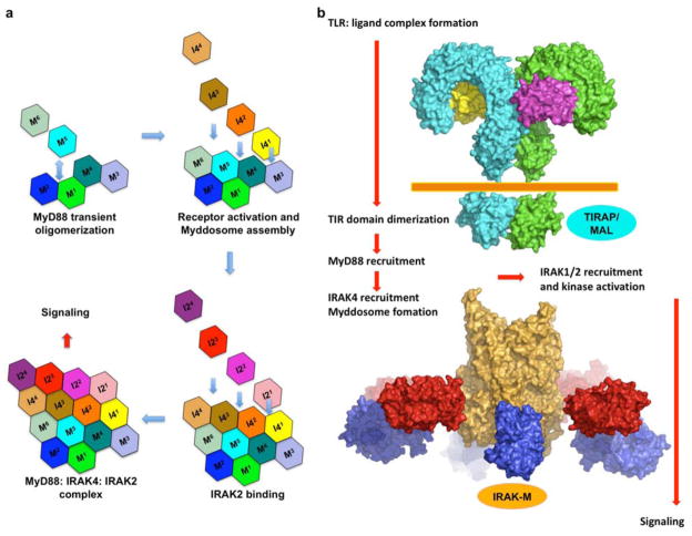
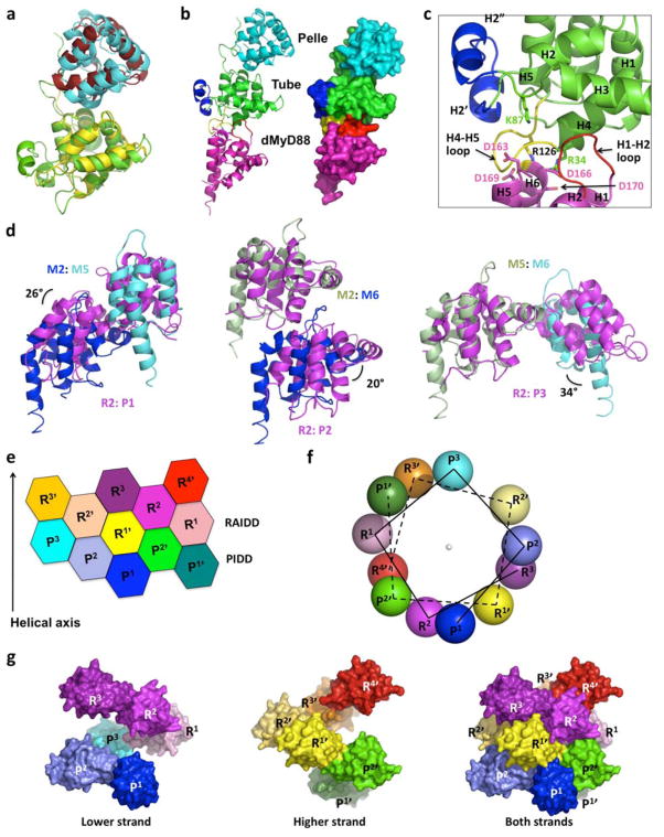
MyD88, IRAK4 and IRAK2 are critical signalling mediators of the TLR/IL1-R superfamily. Here we report the crystal structure of the MyD88-IRAK4-IRAK2 death domain (DD) complex, which surprisingly reveals a left-handed helical oligomer that consists of 6 MyD88, 4 IRAK4 and 4 IRAK2 DDs. Assembly of this helical signalling tower is hierarchical, in which MyD88 recruits IRAK4 and the MyD88-IRAK4 complex recruits the IRAK4 substrates IRAK2 or the related IRAK1. Formation of these Myddosome complexes brings the kinase domains of IRAKs into proximity for phosphorylation and activation. Composite binding sites are required for recruitment of the individual DDs in the complex, which are confirmed by mutagenesis and previously identified signalling mutations. Specificities in Myddosome formation are dictated by both molecular complementarity and correspondence of surface electrostatics. The MyD88-IRAK4-IRAK2 complex provides a template for Toll signalling in Drosophila and an elegant mechanism for versatile assembly and regulation of DD complexes in signal transduction.


Figures

Figure 1
Figure 1. Structure of the ternary Myddosome complex
a, A ribbon diagram of the structure, with the 6 MyD88 molecules in cold colors, the 4 IRAK4 molecules in earth-tone colors and the 4 IRAK2 molecules in warm colors. b, Superposition of MyD88 DD (cyan), IRAK4 DD (yellow) and IRAK2 DD (magenta). Helices H1 to H6 and the H1-H2, H2-H3 and H4-H5 loops are labeled. c, Surface diagram of the complex with each subunit labeled using the same color coding as in a. M: MyD88; I4: IRAK4; I2: IRAK2. d, Planar arrangement of the complex. e, The helical symmetry is shown in a helical wheel representation with each ball representing a molecule and looking down the helical axis. Each molecule is labeled as an integer sequentially from M1 to I24.
Figure 2
Figure 2. Composite interactions and specificity in the ternary complex
a, Schematic representation of a composite binding site. The Nth, (N+1)th, and (N+3)th molecules provide type IIb, Ib and IIIb surfaces to interact with the type IIa, Ia and IIIa surfaces of a downstream molecule, respectively. b, A composite binding site for IRAK4 (yellow) formed from three MyD88 molecules (M3, M4, and M6) through type I, II and III interactions. c: Enlargement of the square in b. Note that the unique H1-H2 loop of MyD88 is critical for Myddosome assembly. d, A composite binding site for IRAK2 (purple) formed from three IRAK4 molecules (I41, I42, and I44) through type I, II, and III interactions. e, Enlargement of the square in d. f, Good charge complementarity between IRAK4 and MyD88 and poor charge complementarity between the top and bottom surfaces of MyD88. g, Good charge complementarity between IRAK2 and IRAK4 and very poor charge complementarity between the top and bottom surfaces of IRAK2.
Figure 3
Figure 3. Model of sequential assembly upon TLR/IL1-R signaling
a, Upon ligand binding, TLR/IL-1Rs, including the cytoplasmic TIR domains, are dimerized or oligomerized. This results in the recruitment of other TIR containing adaptor proteins. In this case, MyD88 is recruited to the receptor complex and the death domain is oligomerized. In the presence of IRAK4, the death domain of IRAK4 (I41) can be recruited to the oligomerized MyD88 DDs through the three interfaces (M3, M4, M6) and quickly forms the binary Myddosome complex. Downstream kinases (IRAK2 in this case, but IRAK1 as well) can then be recruited in a similar fashion and signaling is triggered. b, A model of the TLR receptor signaling complex that recruits the MyD88: IRAK4: IRAK2 complex with proteins drawn in scale. TLRs: cyan and green (PDB code 3FXI for the extracellular domain of TLR4 and PDB code 2J67 for the TIR domain of TLR10). MD2: yellow and magenta (PDB code 3FXI in complex TLR4). Orange: the MyD88: IRAK4: IRAK2 complex. Red: the IRAK4 kinase domain (PDB code 2NRU). Blue: the IRAK2 kinase domain using that of IRAK4.
Figure 4
Figure 4. Common architecture in Drosophila Toll signaling and DD assembly in general
a, Superposition of Tube: Pelle (green and cyan) and IRAK4: IRAK2 (yellow and brick red) complexes. b, A model of the dMyD88: Tube: Pelle complex in both ribbon and surface representations. Pelle is colored in cyan. Tube is colored in green except that the H2-H3 loop insertion (H2′ and H2″) is in blue and the H4-H5 loop is in yellow. The model of dMyD88, obtained based on the MyD88 DD structure but without modeling the insertion in the H1-H2 loop, is colored in magenta except that the H1-H2 loop is in red. c, Enlargement of the dMyD88: Tube interface. Residues important for dMyD88: Tube complex formation are shown in sticks. Note that the real H1-H2 loop of dMyD88 is 6 residues longer than that in the model. d, Superposition of the type I, II, and III interactions in the MyD88: IRAK4: IRAK2 and the PIDD: RAIDD complexes. The resultant angular differences are labeled. e, Planar arrangement of the PIDD: RAIDD complex. R: RAIDD; P: PIDD. Subunits in one strand of the double helix are labeled as R and P while those in the other strand are labeled R′ and P′. f. The helical symmetry is shown in a helical wheel representation with each ball representing a molecule and looking down the helical axis. Note that two helical strands are present in the complex. g, Surface representations of the two helical strands in the PIDD: RAIDD complex. The lower strand is about 5 Å lower than the higher strand, but they can be superimposed well. The lower strand (left) plus the higher strand (middle) equals the PIDD: RAIDD complex (right).

Comment in

References

    1. O’Neill LA. The interleukin-1 receptor/Toll-like receptor superfamily: 10 years of progress. Immunol Rev. 2008;226:10–18. - PubMed
    1. Takeda K, Kaisho T, Akira S. Toll-like receptors. Annu Rev Immunol. 2003;21:335–376. - PubMed
    1. Akira S, Uematsu S, Takeuchi O. Pathogen recognition and innate immunity. Cell. 2006;124:783–801. - PubMed
    1. Beutler B. Inferences, questions and possibilities in Toll-like receptor signalling. Nature. 2004;430:257–263. - PubMed
    1. Verstak B, Hertzog P, Mansell A. Toll-like receptor signalling and the clinical benefits that lie within. Inflamm Res. 2007;56:1–10. - PubMed

Publication types

MeSH terms

Substances