. 2010 May 20;465(7296):373-7.
doi: 10.1038/nature08994.

Myosin II contributes to cell-scale actin network treadmilling through network disassembly

Affiliations

Myosin II contributes to cell-scale actin network treadmilling through network disassembly

Cyrus A Wilson et al. Nature. .

Abstract

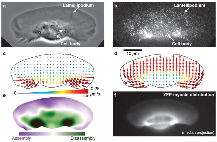
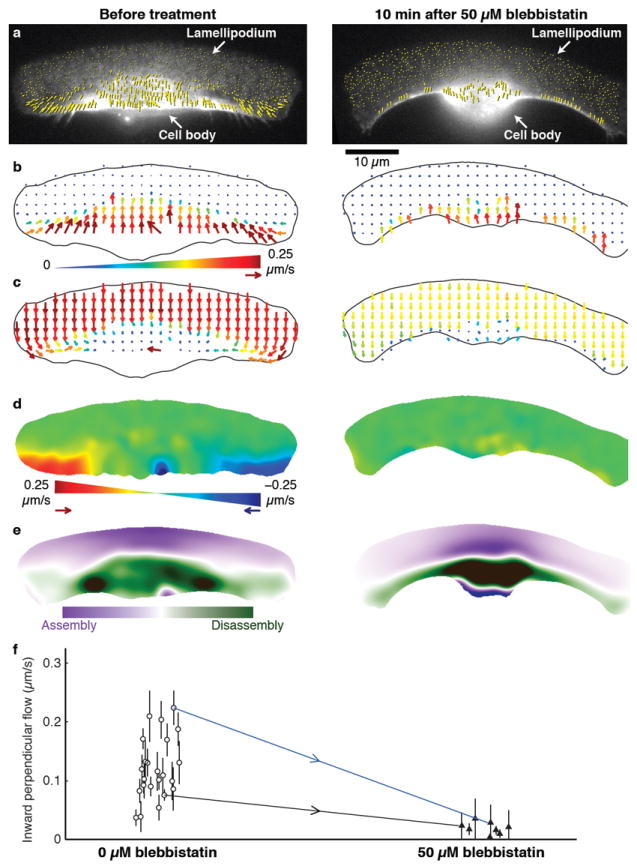
Crawling locomotion of eukaryotic cells is achieved by a process dependent on the actin cytoskeleton: protrusion of the leading edge requires assembly of a network of actin filaments, which must be disassembled at the cell rear for sustained motility. Although ADF/cofilin proteins have been shown to contribute to actin disassembly, it is not clear how activity of these locally acting proteins could be coordinated over the distance scale of the whole cell. Here we show that non-muscle myosin II has a direct role in actin network disassembly in crawling cells. In fish keratocytes undergoing motility, myosin II is concentrated in regions at the rear with high rates of network disassembly. Activation of myosin II by ATP in detergent-extracted cytoskeletons results in rear-localized disassembly of the actin network. Inhibition of myosin II activity and stabilization of actin filaments synergistically impede cell motility, suggesting the existence of two disassembly pathways, one of which requires myosin II activity. Our results establish the importance of myosin II as an enzyme for actin network disassembly; we propose that gradual formation and reorganization of an actomyosin network provides an intrinsic destruction timer, enabling long-range coordination of actin network treadmilling in motile cells.


Figures

Figure 1
Figure 1. Myosin II in keratocytes colocalizes with the primary sites of actin network disassembly
a–e, data and analysis of a single live keratocyte. a, Phase-contrast image of the keratocyte moving upward. b, Fluorescence speckle microscopy (FSM) image of the actin network labeled with a low concentration of phalloidin. c, F-actin flow field based on speckle tracking, in the laboratory frame of reference. Arrow length and color both indicate the speed of actin network flow. d, F-actin flow field in the cell frame of reference. e, Steady-state net F-actin assembly and disassembly. f, Fluorescence image of YFP-tagged myosin regulatory light chain in a keratocyte of similar size and shape to the one shown in a–e. Myosin II is found at low levels throughout the lamellipodium and at the highest concentrations in two foci flanking the cell body, which coincide with the primary sites of actin network disassembly as shown in e.
Figure 2
Figure 2. Inhibition of myosin II blocks inward flow and alters the pattern of disassembly of the actin network
a–e, Analysis of actin network flow in a single keratocyte before (left) and ~10 min after (right) addition of 50 μM blebbistatin. a, Raw F-actin speckle flow measurements (yellow arrows) in the laboratory frame of reference, superimposed on the corresponding FSM frames. b, Resampled flow field in the laboratory frame of reference. c, Resampled flow field in the cell frame of reference. d, Maps showing the component of network flow perpendicular to the direction of cell movement (“perpendicular flow”), in the cell frame of reference. Red indicates F-actin flow toward the right; blue, toward the left. Actin network movement in the green regions is parallel to the direction of cell locomotion. Blebbistatin treatment abolishes inward flow. e, Steady-state assembly/disassembly maps. Before blebbistatin treatment, the highest rate of disassembly is found in two foci flanking the cell body. After blebbistatin treatment, disassembly is distributed along the rear margin. f, Inward perpendicular flow of the actin network in untreated (white circles; n = 23) and blebbistatin-treated (black triangles; n = 8) cells. Error bars indicate average ± s.d. over each movie (typically 4 min). Measurements were made both before and after treatment for two of the cells; blue and black arrows connect the corresponding data points. The data points connected by the blue arrow correspond to the cell shown in a–e. Compare Supplementary Movie 1.
Figure 3
Figure 3. Jasplakinolide specifically halts actin dynamics of cells in which myosin II is inhibited
a, Raw F-actin speckle flow measurements in the cell frame of reference (yellow arrows) superimposed on the corresponding FSM frames, for a cell in 50 μM blebbistatin before (top) and ~2 min after (middle) addition of 1 μM jasplakinolide. Bottom, a separate cell in jasplakinolide alone. b, F-actin flow magnitude maps corresponding to a. The combination of blebbistatin and jasplakinolide immobilizes the actin network, an effect that is not achieved by either drug alone. c–e, Fixed, phalloidin-labeled keratocytes, untreated (c) or treated with 50 μM blebbistatin (d) or 1 μM jasplakinolide (e). Consistent with impaired actin network disassembly, blebbistatin-treated cells accumulate F-actin along the rear margin; jasplakinolide-treated cells underneath the cell body. f, Treatment with either 5 nM latrunculin A, 50 μM blebbistatin, or 1 μM jasplakinolide can slow cells relative to the control population. The combination of blebbistatin and latrunculin A or jasplakinolide and latrunculin A has no significant further effect on cell speed than either drug alone. The combination of blebbistatin and jasplakinolide significantly (p < 0.05 by Tukey’s test) and synergistically slows cell locomotion. g, Blebbistatin causes significant (p < 0.05 by Tukey’s test) F-actin accumulation in the trailing “tails” (but not in the cell body), whereas jasplakinolide causes accumulation underneath the cell body (but not in the “tails”), relative to untreated cells. a.u., arbitrary units (see Methods). Compare c–e. Box-and-whisker plots (f and g) indicate the mean, 95% confidence interval (C.I.), extrema and quartiles, for the indicated number of cells (n) in each treatment group. Compare Supplementary Movie 3.
Figure 4
Figure 4. Actin network disassembly in the rear of detergent-extracted keratocyte cytoskeletons is ATP-dependent and blebbistatin-sensitive, consistent with a direct role for myosin II in this process
a–d, ATP triggers an acute loss of actin network in the rear region of the cell, where myosin II is localized (compare Fig. 1f). a, A detergent-extracted and phalloidin-labeled keratocyte cytoskeleton. b, The same cytoskeleton 7 min after addition of 1 mM ATP. c, Overlay of initial frame (a, cyan) and frame at 7 min (b, yellow); regions with increase, decrease, or no change in net intensity appear yellow, cyan, or white, respectively. d, Time evolution of fluorescence intensities (normalized at t = 0) in the indicated regions. Time points for a mock buffer wash (chevron) and ATP addition (black arrowhead) are indicated. e–h, In a cell treated with 50 μM blebbistatin for 30 min prior to extraction, addition of ATP does not induce a loss of actin network. There is a slow loss of fluorescence due to photobleaching or background dissociation. i–l, The F-actin severing protein villin rapidly disassembles the lamellipodial actin network, demonstrating that this part of the cytoskeleton is not protected against a general disassembling activity. 0.1 μM GST-villin was added instead of ATP (arrow in l). m–t, Addition of GST-villin (arrows in p, t) in addition to ATP (arrowheads in p, t), in either order, results in complete, rapid disassembly of the actin network. Compare Supplementary Movie 4.

References

    1. Abercrombie M. The Croonian lecture, 1978: the crawling movement of metazoan cells. Proc R Soc Lond B Biol Sci. 1980;207:129–47.
    1. Theriot JA, Mitchison TJ. Actin microfilament dynamics in locomoting cells. Nature. 1991;352:126–31. - PubMed
    1. Pollard TD, Borisy GG. Cellular motility driven by assembly and disassembly of actin filaments. Cell. 2003;112:453–65. - PubMed
    1. Euteneuer U, Schliwa M. Persistent, directional motility of cells and cytoplasmic fragments in the absence of microtubules. Nature. 1984;310:58–61. - PubMed
    1. Small JV, Herzog M, Häner M, Abei U. Visualization of actin filaments in keratocyte lamellipodia: negative staining compared with freeze-drying. J Struct Biol. 1994;113:135–41. - PubMed

Publication types