. 2010 May 11;5(5):e10580.
doi: 10.1371/journal.pone.0010580.

Interdomain interactions control Ca2+-dependent potentiation in the cation channel TRPV4

Affiliations

Interdomain interactions control Ca2+-dependent potentiation in the cation channel TRPV4

Rainer Strotmann et al. PLoS One. .

Abstract

Several Ca(2+)-permeable channels, including the non-selective cation channel TRPV4, are subject to Ca(2+)-dependent facilitation. Although it has been clearly demonstrated in functional experiments that calmodulin (CaM) binding to intracellular domains of TRP channels is involved in this process, the molecular mechanism remains elusive. In this study, we provide experimental evidence for a comprehensive molecular model that explains Ca(2+)-dependent facilitation of TRPV4. In the resting state, an intracellular domain from the channel N terminus forms an autoinhibitory complex with a C-terminal domain that includes a high-affinity CaM binding site. CaM binding, secondary to rises in intracellular Ca(2+), displaces the N-terminal domain which may then form a homologous interaction with an identical domain from a second subunit. This represents a novel potentiation mechanism that may also be relevant in other Ca(2+)-permeable channels.


Conflict of interest statement

Competing Interests: The authors have declared that no competing interests exist.

Figures

Figure 1
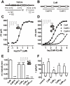
Figure 1. Lobe-specific CaM binding to TRPV4-C2.
(A) TRPV4 topology. Ankyrin domains, transmembrane helices and the C-terminal CaM binding site associated with Ca2+-dependent potentiation are shown as black, white and grey boxes in the linearized TRPV4 structure. The positions of the protein fragments, peptides P1–5 and the N-terminal mutations (asterisk) used in this study are indicated below. For the precise positions see Table S1 (B) Structure of the CaM molecule. The EF hand domains 1 to 4 are shown as white boxes and the positions of the homologous lobe fragments used for the interaction experiments are indicated. (C) Ca2+-dependence of CaM binding to the fluorophore-labeled P5 peptide measured by fluorescence polarization. (D) Binding of the individual CaM lobes to fluorophore-labeled P5 peptide at 100 µM Ca2+. (E) Binding to the C2 fragment of the CaM lobes or CaM mutants that are lobe-specifically (CaM12, CaM34) or fully (CaM1234) Ca2+ binding-deficient (4 independent experiments, p≤0.001). (F) In HEK293 cells coexpressing TRPV4 and the indicated CaM mutants, whole-cell currents were activated by the application of 4α-PMA in Ca2+-free bath solutions. Current increases at +100 mV and −100 mV were measured after addition of 2 mM Ca2+ and compared to CaM overexpression (p = 0.013 and 0.007, respectively, for CaM12).
Figure 2
Figure 2. AlphaScreen-based CaM interaction assay.
Optical stimulation of phthalocyanine compounds contained in the AlphaScreen donor beads excites ambient oxygen molecules into the metastable singlet state. The energy conveyed by the molecule is converted into light emission by the acceptor bead. Owing to the limited lifespan of singlet oxygen in solution, the AlphaScreen signal emission is highly dependent on the distance between the two beads. In this study, AlphaScreen donor and acceptor beads were modified for the detection of CaM/TRPV4 or interdomain interactions. Streptavidin-coated donor and anti-Flag acceptor beads were used to attach the proteins to the bead surface.
Figure 3
Figure 3. The C2 fragment interacts with an N-terminal domain.
(A) P2 and CaM bind competitively to C2. C2-Flag interaction with P2-biotin (squares) or, as control protein, C2-biotin (circles) was measured in an AlphaScreen interaction assay in the presence of CaM at the indicated concentrations or in the absence of CaM (control). Buffers contained either 100 µM Ca2+ (filled symbols) or 1 mM EGTA (open symbols). A strong constitutive interaction between C2 and P2 was observed that was inhibited by Ca2+/CaM. (B) The C2-P2 interaction is Ca2+-independent over a wide range of Ca2+ concentrations. (C and D) Mutation of specific amino acids in the N-terminal binding domain abolishes the interdomain interaction. Binding of C2-biotin to the N2-Flag (wt) or the mutants N2-WIA and N2-KRA given in D was measured at concentrations between 10 pM and 1 µM in buffers containing 100 µM Ca2+. (E) Curve maxima from C were tested against wild type. Both mutants showed significantly lower interaction signals (p<0.0001, 5 independent experiments).
Figure 4
Figure 4. The TRPV4 N and C termini interact in HEK293 cells.
(A) YFP-fused C1 fragment and CFP-fused N1 (left) or CFP (right) were coexpressed in HEK293 cells and the donor and acceptor fluorescences (black and grey traces, respectively) measured during selective YFP photobleaching (bar, 6 and 3 independent experiments). (B) FRET acceptor bleaching measurements as shown in A were performed with the indicated donor/acceptor combinations. YFP and CFP, respectively, were used as acceptor and donor controls (3 independent experiments).
Figure 5
Figure 5. Calcium-dependent interdomain interaction in TRPV4.
(A) A CFP-TRPV4-YFP fusion construct (TRPV4) or TRPV4-YFP cotransfected with CFP (control) were subjected to FRET acceptor bleaching experiments after expression in HEK293 cells. (B) FRET efficiencies were calculated by the extrapolated donor increase at full acceptor photobleaching (three cells shown from each group). The FRET efficiencies from the experiments shown in (A) (labeled C-V4-Y and control), and cells cotransfected with CFP-TRPV4 and TRPV4-YFP (labeled C−V4+V4−Y), were 14.6±1.1, 2.0±0.8 and 19.6±1.9. Both CFP-TRPV4-YFP and CFP-TRPV4+TRPV4-YFP interactions were significantly different from control (inset, p<0.0001, n = 7, 20 and 13, respectively). (C) The donor and FRET channels were recorded in HEK293 cells expressing CFP-TRPV4-YFP during application of hypotonic medium and 10 µM carbachol.
Figure 6
Figure 6. TRPV4 mutations that disrupt the interaction between the N- and C-terminal domains result in channels that are strongly activated in the absence of extracellular Ca2+.
(A–C) Time courses (left) of currents at -100 and +100 mV showing the activation by 4α-PMA (1 µM) in a nominally Ca2+-free solution (0 Ca2+) and the effect of readdition of 2 mM Ca2+ for wt TRPV4 (A) and the mutants WIA (B) and KRA (C). The IV-relationships (right) were recorded in 4α-PMA just before and shortly after switching to 2 mM Ca2+ as indicated by the arrows. (D) Mean current densities at +100 and −100 mV measured after break-in (basal) and at the start of 4α-PMA application in a nominally Ca2+-free solution (0 Ca2+), and the mean maximum inward and outward current densities attained in 4α-PMA in the nominally Ca2+-free solution, and following the addition of 2 mM Ca2+ in 4α-PMA (see numbers in A). The currents measured after stimulation in nominally Ca2+-free solutions at -100 and +100 mV for the WIA (p = 0.014 and 0.023) and KRA mutants (p = 0.012 and 0.007) were significantly larger than the wild type (n = 9, 8 and 5 for wt, WIA and KRA).
Figure 7
Figure 7. The P2 peptide contributes to homodimerization of the TRPV4 N terminus.
(A) The purified and biotinylated N2-GST protein was subjected to SDS-PAGE, blotted and visualized in avidin-peroxidase overlay experiments. In addition to the monomeric form at 34.8 kD, a second band at a size consistent with that of the N2 dimer was detected. (B) The P2 peptide interacts with the wild type N2 fragment and the WIA but not the KRA mutant. In an AlphaScreen experiment, biotinylated P2 peptide was subjected to interaction with wild type N2 fragment (wt) or the mutants WIA or KRA at the indicated concentrations. (C) The full-length TRPV4 N termini show dimerization in FRET acceptor bleaching experiments. (p<0.0001, 4 and 3 independent experiments). (D) The interaction between P2-biotin and C2-Flag is inhibited by the C2 fragment with a 3.5-fold lower IC50 than the N2 fragment. (E) The complex between CaM-biotin and C2-Flag at 100 µM Ca2+ is inhibited by C2 with a 14-fold higher affinity than by N2.
Figure 8
Figure 8. Proposed TRPV4 potentiation mechanism.
(A) In the resting state, an N-terminal site (filled box) binds to a C-terminal domain (open box) thus permitting only weak channel activation. The hatched lines in the fourth transmembrane region are meant to indicate that the interaction can occur between channel subunits or possibly within a channel subunit. (B) Increase of the intracellular calcium concentration to micromolar concentrations leads to Ca2+/CaM binding to the C-terminal site and displacement of the N-terminal interaction site. The corresponding conformational change induces further increase in channel activity. In the potentiated state, the N-terminal binding site may form a homologous interaction with a second channel subunit.

Similar articles

Cited by

References

    1. Zhang Z, Tang J, Tikunova S, Johnson JD, Chen Z, et al. Activation of Trp3 by inositol 1,4,5-trisphosphate receptors through displacement of inhibitory calmodulin from a common binding domain. Proc Natl Acad Sci U S A. 2001;98:3168–3173. - PMC - PubMed
    1. Singh BB, Liu X, Tang J, Zhu MX, Ambudkar IS. Calmodulin regulates Ca(2+)-dependent feedback inhibition of store-operated Ca(2+) influx by interaction with a site in the C terminus of TrpC1. Mol Cell. 2002;9:739–750. - PubMed
    1. Numazaki M, Tominaga T, Takeuchi K, Murayama N, Toyooka H, et al. Structural determinant of TRPV1 desensitization interacts with calmodulin. Proc Natl Acad Sci U S A. 2003;100:8002–8006. - PMC - PubMed
    1. Niemeyer BA, Bergs C, Wissenbach U, Flockerzi V, Trost C. Competitive regulation of CaT-like-mediated Ca2+ entry by protein kinase C and calmodulin. Proc Natl Acad Sci U S A. 2001;98:3600–3605. - PMC - PubMed
    1. Tang J, Lin Y, Zhang Z, Tikunova S, Birnbaumer L, et al. Identification of common binding sites for calmodulin and inositol 1,4,5-trisphosphate receptors on the carboxyl termini of trp channels. J Biol Chem. 2001;276:21303–21310. - PMC - PubMed

Publication types