. 2010 May 11;5(5):e10570.
doi: 10.1371/journal.pone.0010570.

Antiretroviral treatment start-time during primary SIV(mac) infection in macaques exerts a different impact on early viral replication and dissemination

Affiliations

Antiretroviral treatment start-time during primary SIV(mac) infection in macaques exerts a different impact on early viral replication and dissemination

Pierre Sellier et al. PLoS One. .

Abstract

Background: The time of infection is rarely known in human cases; thus, the effects of delaying the initiation of antiretroviral therapy (ART) on the peripheral viral load and the establishment of viral reservoirs are poorly understood.

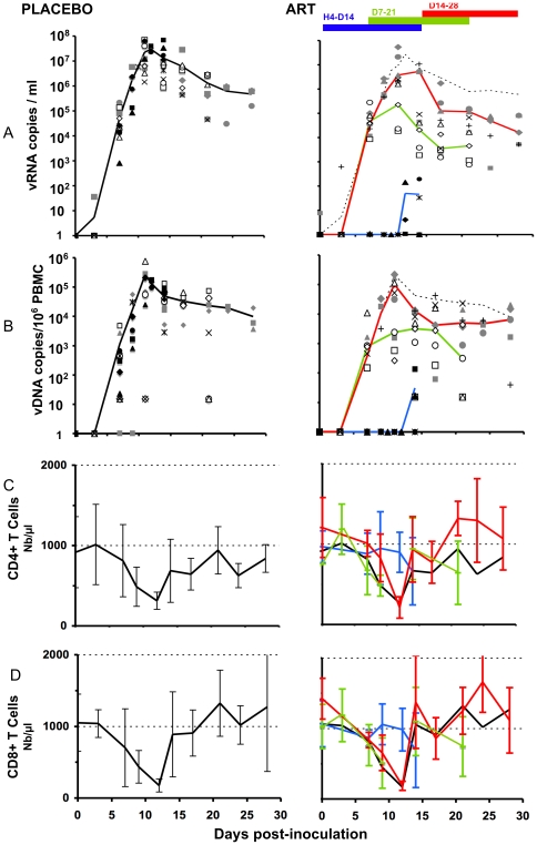
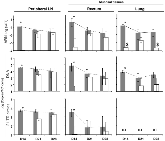
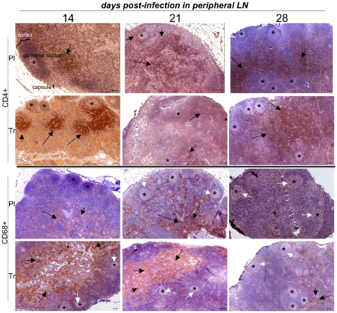
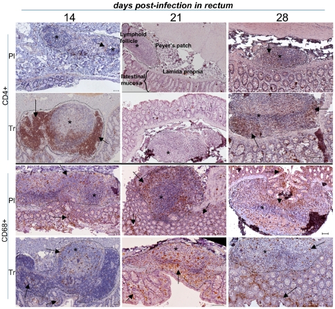
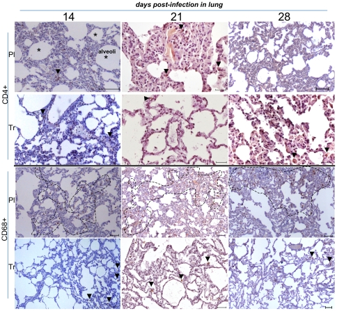
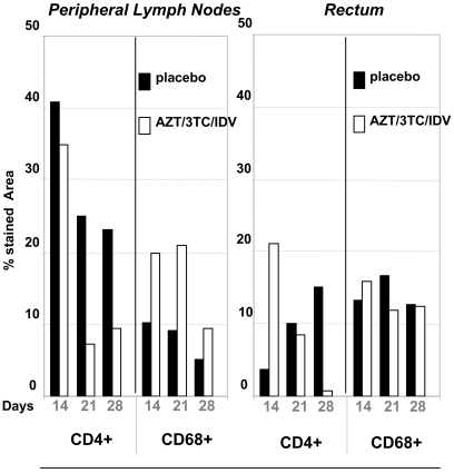
Methodology/principal findings: Six groups of macaques, infected intravenously with SIV(mac251), were given placebo or antiretroviral therapy to explore reservoir establishment; macaques were treated for 2 weeks, with treatment starting 4 hours, 7 or 14 days after infection. Viral replication and dissemination were measured in the gut (rectum), in the lung and in blood and lymphoid tissues (peripheral lymph nodes), by quantifying viral RNA, DNA and 2LTR circles. We used immunohistochemistry (CD4 and CD68) to assess the impact of these treatments on the relative amount of virus target cells in tissue. Treatment that was started 4 hours post-infection (pi) decreased viral replication and dissemination in blood and tissue samples, which were assessed on day 14 (RNA/DNA/2LTR circles). The virus remained detectable and lymphoid tissues were activated in LN and the gut in both placebo- and ART-treated animals. Viral RNA in plasma continued to be lower in macaques treated seven days after infection; however, this was not the case for viral DNA in peripheral blood mononuclear cells. There was a small but significant difference in RNA and DNA levels in tissues between placebo- and ART-treated animals on day 21. When started 14 days after infection, treatment resulted in a limited decrease in the plasma viral load.

Conclusions: Treatment that was started 4 hours after infection significantly reduced viral replication and dissemination. When started 7 days after infection, it was of slight virological benefit in peripheral blood and in tissues, and treatment was even less effective if started 14 days pi. These data favor starting ART no longer than one week after intravenous SIV(mac251) exposure.


Conflict of interest statement

Competing Interests: The authors have declared that no competing interests exist.

Figures

Figure 1
Figure 1. Peripheral viral loads and changes in CD4, and CD8 T cells in placebo- and ART-treated animals.
Placebo animals are shown on the left side (median in dark line), and treated animals, on the right side of the figure. Values for animals are given as a filled symbol and a star (group H4-D14); open symbol and x (group D7-21); grey symbols and + (group D14-28). Median curves are shown according to the date of treatment initiation, with the black or dotted line for placebo and blue, green, and red lines for the H4-D14, D7-21 and D14-28 groups, respectively. A) Plasma viral load; vRNA copies/ml of plasma. B) Total viral DNA in PBMCs; vDNA copies/106 PBMC. C) CD4 changes in absolute number (mean±SD) D) CD8 changes in absolute number (mean±SD).
Figure 2
Figure 2. Change in viral loads in tissues in animals given placebo or treatment over the course of 14 days.
Viral loads were expressed using RNA Log (ΔCT), DNA and 2LTR Log(Copies/106 cells) at the top, middle and at the bottom of the figure, in peripheral LN, rectum, and lung (left side, middle, and right side), respectively. LN: lymph nodes; placebo animals (Gray bars), treated animals (white bars). Vertical bars represented the 95% confidence interval (95% CI). 2LTR values in the lung were below the threshold and were listed as BT. $ viral RNA was detected in only 1 animal in ART-treated animals on day 14 and day 28. * Significant differences between placebo and treated animals.
Figure 3
Figure 3. Low magnification of IHC stains of peripheral LN samples in placebo- and ART-treated animals.
For clarity, we identified the germinal center (GC), cortex and capsule from a lymph node in one of the panels. The limit of the white pulp region is shown by a dotted line, and germinal centers are shown using stars. CD4+ staining surrounded lymphoid follicles (black arrows), whereas CD68+ staining was mainly localized in the white pulp (black arrows) and to a lesser extent in the GC (white arrows). Target cells were shown at the top (CD4+) and the bottom (CD68+) of the figure. For each pair of presented animals, placebo-treated animals were located above, and PEP-treated animals below. Animals killed on day 14, day 21, and day 28 were shown from the left to right of the figure. The horizontal bar on each panel corresponds to 100 µm. Each picture was representative of explored tissues and was cropped from a large image. Lymph node and GALT architecture organization is shown; specific staining is colored brown.
Figure 4
Figure 4. Low magnification of IHC stains of rectal samples in placebo- and ART-treated animals.
The lymphoid-rich area (Payer's patch) was sectioned by a dotted line and stars indicated lymphoid follicles; rare CD4+ lymphocyte staining is highlighted by arrows. CD68+ staining is seen in both lymphoid follicles and interstitial zones (arrows). Presentation as in Figure 3.
Figure 5
Figure 5. Low magnification of IHC stains of lung samples in placebo- and ART-treated animals.
CD4+ staining was occasionally found in the lung (arrow). CD68+ (alveolar monocyte/macrophages) staining was greater in the placebo (dotted line) than in ART-treated animals (arrow). A random portion of the lung was taken from a large image. Presentation as in Figure 3.
Figure 6
Figure 6. Percentage of the area stained for CD4+ and CD68+ cells (IHC) in peripheral LN and rectum, according to treatment groups and dates of initiation.
Dark dot blots symbolized placebo-treated animals, and white dot blots, PEP-treated animals. The percentage of the stained area was determined by a quantification performed using two animals per group (see Material and methods).

Similar articles

Cited by

References

    1. Smith DK, Grohskopf LA, Black RJ, Auerbach JD, Veronese F, et al. Antiretroviral postexposure prophylaxis after sexual, injection-drug use, or other nonoccupational exposure to HIV in the United States: recommendations from the U.S. Department of Health and Human Services. MMWR Recomm Rep. 2005;54:1–20. - PubMed
    1. Yeni P. Paris: Medecine-Sciences Flammarion; 2008. Prise en charge medicale des personnes infectees par le VIH.412. Recommandations du groupe d'experts.
    1. Otten RA, Smith DK, Adams DR, Pullium JK, Jackson E, et al. Efficacy of postexposure prophylaxis after intravaginal exposure of pig-tailed macaques to a human-derived retrovirus (human immunodeficiency virus type 2). J Virol. 2000;74:9771–9775. - PMC - PubMed
    1. Tsai CC, Emau P, Follis KE, Beck TW, Benveniste RE, et al. Effectiveness of postinoculation (R)-9-(2-phosphonylmethoxypropyl) adenine treatment for prevention of persistent simian immunodeficiency virus SIVmne infection depends critically on timing of initiation and duration of treatment. J Virol. 1998;72:4265–4273. - PMC - PubMed
    1. Merchant RC, Keshavarz R, Low C. HIV post-exposure prophylaxis provided at an urban paediatric emergency department to female adolescents after sexual assault. Emerg Med J. 2004;21:449–451. - PMC - PubMed

Publication types

MeSH terms

Substances