. 2010 May 11;5(5):e10549.
doi: 10.1371/journal.pone.0010549.

Antibodies targeted to the brain with image-guided focused ultrasound reduces amyloid-beta plaque load in the TgCRND8 mouse model of Alzheimer's disease

Affiliations

Antibodies targeted to the brain with image-guided focused ultrasound reduces amyloid-beta plaque load in the TgCRND8 mouse model of Alzheimer's disease

Jessica F Jordão et al. PLoS One. .

Abstract

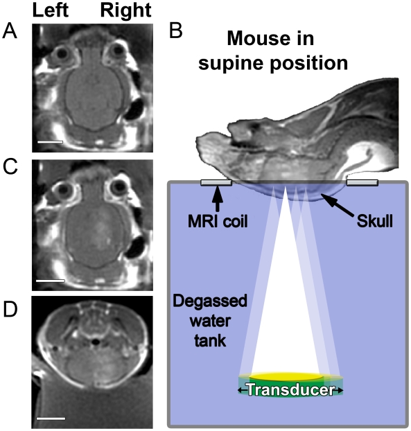
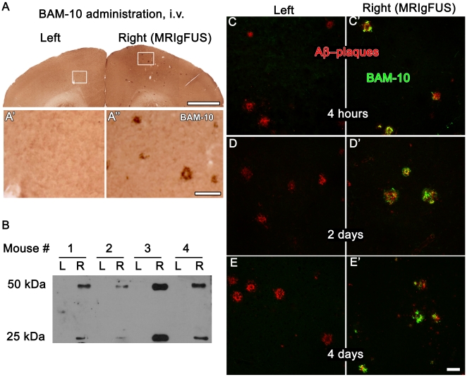
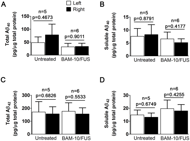
Immunotherapy for Alzheimer's disease (AD) relies on antibodies directed against toxic amyloid-beta peptide (Abeta), which circulate in the bloodstream and remove Abeta from the brain. In mouse models of AD, the administration of anti-Abeta antibodies directly into the brain, in comparison to the bloodstream, was shown to be more efficient at reducing Abeta plaque pathology. Therefore, delivering anti-Abeta antibodies to the brain of AD patients may also improve treatment efficiency. Transcranial focused ultrasound (FUS) is known to transiently-enhance the permeability of the blood-brain barrier (BBB), allowing intravenously administered therapeutics to enter the brain. Our goal was to establish that anti-Abeta antibodies delivered to the brain using magnetic resonance imaging-guided FUS (MRIgFUS) can reduce plaque pathology. To test this, TgCRND8 mice received intravenous injections of MRI and FUS contrast agents, as well as anti-Abeta antibody, BAM-10. MRIgFUS was then applied transcranially. Within minutes, the MRI contrast agent entered the brain, and BAM-10 was later found bound to Abeta plaques in targeted cortical areas. Four days post-treatment, Abeta pathology was significantly reduced in TgCRND8 mice. In conclusion, this is the first report to demonstrate that MRIgFUS delivery of anti-Abeta antibodies provides the combined advantages of using a low dose of antibody and rapidly reducing plaque pathology.


Conflict of interest statement

Competing Interests: The authors have declared that no competing interests exist.

Figures

Figure 1
Figure 1. Magnetic resonance imaging-guided focused ultrasound (MRIgFUS) increases the permeability of the blood brain barrier (BBB).
T1-weighted contrast-enhanced MRI scans were used to position ultrasound foci prior to treatment (A) and following transcranial FUS (B–D), were used to monitor the entry of gadolinium in the brain. Mice were positioned in a supine position (B) and injected in the tail vein with microbubbles and gadolinium while ultrasound was applied to 4 aligned spots on the right hemisphere of the brain. Increased BBB permeability was monitored by MRI, visualizing contrast enhancement by the influx of gadolinium (B–D). [A–D: 128×128, TE/TR = 10.4/500.0, FOV = 4cm, Slice 1mm, ETL = 4, 3NEX]. Scale bars: A, C, D = 0.5 cm.
Figure 2
Figure 2. BAM-10 entered the right, MRIgFUS-treated, side of the brain and bound to Aβ plaques.
Following intravenous (i.v.) administration of biotinylated BAM-10 and treatment with MRIgFUS, the entry of biotinylated BAM-10 into the brain of TgCRND8 mice was evaluated by processing brain sections with streptavidin-horseradish peroxidase and standard histochemistry procedures (A). Biotinylated BAM-10 was not found on left side of the brain (A′). In contrast, the right side of the brain, exposed to FUS, had abundant biotinylated BAM-10-deposits (A″). Biochemical analysis of brain tissue by immunoprecipitation and western blotting (B, 4 mice represented) also showed that biotinylated BAM-10 was not detected on the left (B, L) and abundant on the right (B, R) side of the brain, which received MRIgFUS. Brain sections from mice sacrificed at 4 hours (C, C′), 2 days (D, D′) and 4 days (E, E′) post-treatment were double stained with streptavidin-Alexa 488 to detect biotinylated BAM-10 (green) and a rabbit Aβ42 antibody conjugated with Cy3 (red). Biotinylated BAM-10 was present at all time points on the right side of the brain receiving MRIgFUS and it co-localized with Aβ-positive plaques. Scale bars: A = 1 mm; A′, A″ = 100 µm; C-E′ = 50 µm.
Figure 3
Figure 3. Focused ultrasound delivery of BAM-10 to the brain reduces Aβ plaque pathology in TgCRND8 mice.
Coronal sections were stained with streptavadin-Cy2 for (A) biotinylated BAM-10 and (B) anti-Aβ antibody 6F3D for plaques to demonstrate that 6F3D binds to plaques which are strongly (arrows) and weakly (arrowheads) positive for BAM-10 (C). Once it was confirmed that BAM-10 does not interfere with 6F3D plaque detection, sections for mice in each treatment group were stained with 6F3D and the stereology software was used to draw contours outlining the FUS-targeted region (determined from MRI post-treatment scans) on the right side of the brain and an equivalent region on the contralateral side (D). Plaques were counted and measured at high magnification (E), using stereological methods. In 4 days, the mean (F) count, (G) size and (H) surface area of Aβ plaques on the right, MRIgFUS-targeted side of the brain was consistently reduced in comparison to the left side of the brain only for the BAM-10/FUS-treated mice (F–H; n = 6, paired t-tests, p = 0.008, p = 0.048 and p = 0.003, respectively). This difference is unique to BAM-10/FUS treatment as there was no significant difference between right and left side of the brain in mice from other treatment groups (F′–H′: BAM-10 treated group, n = 6, paired t-tests, p = 0.294, p = 0.941 and p = 0.402; F″–H″: Untreated group, n = 6, paired t-tests, p = 0.502, p = 0.690, p = 0.610). Scale bars: A–C = 100 µm (inset = 20 µm); D = 1 mm; E = 50 µm.
Figure 4
Figure 4. Aβ levels in TgCRND8 mice after one BAM-10/FUS treatment.
Cortical homogenates were analyzed for total Aβ40 (A), soluble Aβ40 (B), total Aβ42 (C) and soluble Aβ42 (D). There was no significant difference detected any Aβ species and fractions between the left and right hemispheres.

Similar articles

Cited by

References

    1. Weiner HL, Frenkel D. Immunology and immunotherapy of Alzheimer's disease. Nat Rev Immunol. 2006;6:404–416. - PubMed
    1. Hawkes CA, McLaurin J. Immunotherapy as treatment for Alzheimer's disease. Expert Rev Neurother. 2007;7:1535–1548. - PubMed
    1. Thakker DR, Weatherspoon MR, Harrison J, Keene TE, Lane DS, et al. Intracerebroventricular amyloid-beta antibodies reduce cerebral amyloid angiopathy and associated micro-hemorrhages in aged Tg2576 mice. Proc Natl Acad Sci U S A. 2009;106:4501–4506. - PMC - PubMed
    1. Kotilinek LA, Bacskai B, Westerman M, Kawarabayashi T, Younkin L, et al. Reversible memory loss in a mouse transgenic model of Alzheimer's disease. J Neurosci. 2002;22:6331–6335. - PMC - PubMed
    1. Hynynen K, McDannold N, Vykhodtseva N, Jolesz FA. Noninvasive MR imaging-guided focal opening of the blood-brain barrier in rabbits. Radiology. 2001;220:640–646. - PubMed

Publication types

MeSH terms