. 2010 May 11;5(5):e10575.
doi: 10.1371/journal.pone.0010575.

Hepatitis C virus controls interferon production through PKR activation

Affiliations

Hepatitis C virus controls interferon production through PKR activation

Noëlla Arnaud et al. PLoS One. .

Abstract

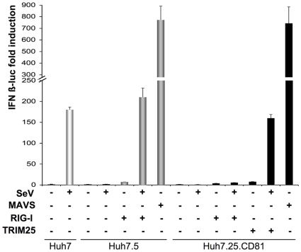
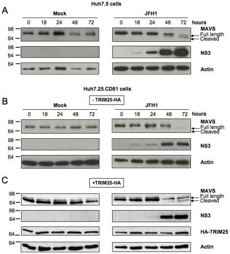
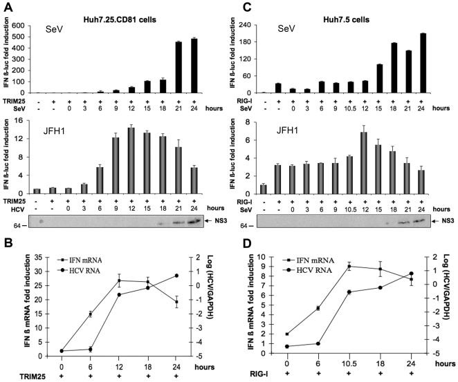
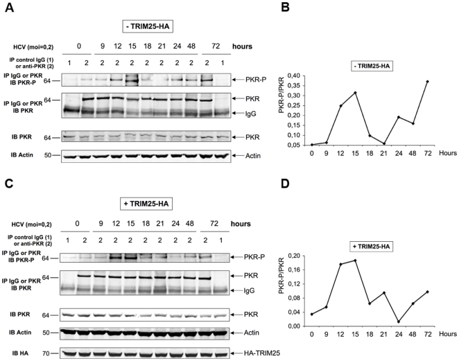
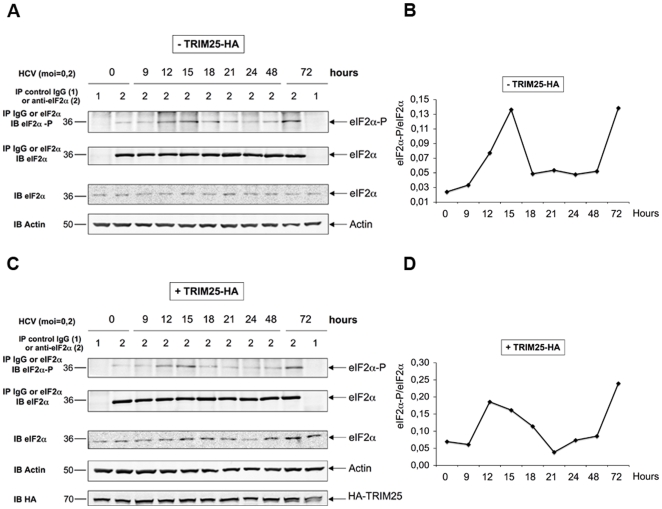
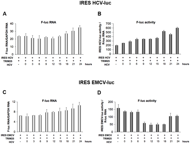
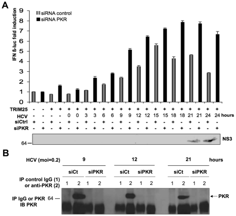
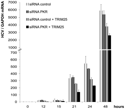
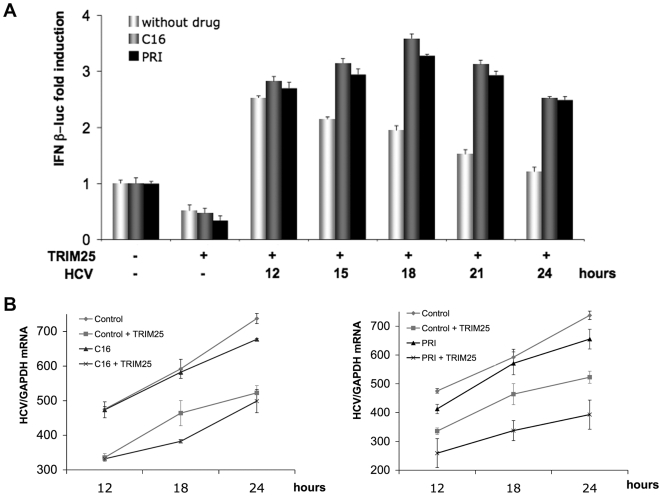
Hepatitis C virus is a poor inducer of interferon (IFN), although its structured viral RNA can bind the RNA helicase RIG-I, and activate the IFN-induction pathway. Low IFN induction has been attributed to HCV NS3/4A protease-mediated cleavage of the mitochondria-adapter MAVS. Here, we have investigated the early events of IFN induction upon HCV infection, using the cell-cultured HCV JFH1 strain and the new HCV-permissive hepatoma-derived Huh7.25.CD81 cell subclone. These cells depend on ectopic expression of the RIG-I ubiquitinating enzyme TRIM25 to induce IFN through the RIG-I/MAVS pathway. We observed induction of IFN during the first 12 hrs of HCV infection, after which a decline occurred which was more abrupt at the protein than at the RNA level, revealing a novel HCV-mediated control of IFN induction at the level of translation. The cellular protein kinase PKR is an important regulator of translation, through the phosphorylation of its substrate the eIF2alpha initiation factor. A comparison of the expression of luciferase placed under the control of an eIF2alpha-dependent (IRES(EMCV)) or independent (IRES(HCV)) RNA showed a specific HCV-mediated inhibition of eIF2alpha-dependent translation. We demonstrated that HCV infection triggers the phosphorylation of both PKR and eIF2alpha at 12 and 15 hrs post-infection. PKR silencing, as well as treatment with PKR pharmacological inhibitors, restored IFN induction in JFH1-infected cells, at least until 18 hrs post-infection, at which time a decrease in IFN expression could be attributed to NS3/4A-mediated MAVS cleavage. Importantly, both PKR silencing and PKR inhibitors led to inhibition of HCV yields in cells that express functional RIG-I/MAVS. In conclusion, here we provide the first evidence that HCV uses PKR to restrain its ability to induce IFN through the RIG-I/MAVS pathway. This opens up new possibilities to assay PKR chemical inhibitors for their potential to boost innate immunity in HCV infection.


Conflict of interest statement

Competing Interests: The authors have declared that no competing interests exist.

Figures

Figure 1
Figure 1. Ectopic expression of TRIM25 restores IFN induction in Huh7.25.CD81 cells.
Huh7, Huh7.5 and Huh7.25.CD81 cells were transfected with the pGL2-IFNβ-FLUC/pRL-TK-RLUC reporter plasmids alone or the in presence of plasmids expressing RIG-I (200 ng), TRIM25 (100 ng) or MAVS (400 ng). 24 hrs post transfection, cells were mock-infected or infected with Sendaï virus (SeV) (40 HAU/ml). 24 hrs after infection, luciferase activity was measured and F-luc was normalized against R-luc. IFN expression was expressed as fold induction over control cells that were simply transfected with pGL2-IFNβ-FLUC/pRL-TK-RLUC. Error bars represent the mean ± S.D. for triplicates.
Figure 2
Figure 2. Kinetics of MAVS cleavage in Huh7.25.CD81 cells after JFH1 infection.
Huh7.5 cells and Huh7.25.CD81 cells were transfected with an HA-TRIM25 expressing plasmid or with an empty plasmid. 24 hrs post-transfection, cells were mock-infected or infected with JFH1 (m.o.i = 0.05) for the indicated times and cell lysates were generated. Cell extracts (50 µg) were subjected to SDS-12.5% PAGE and blotted with anti-MAVS, anti-NS3, anti-HA or anti-actin as indicated. The arrows indicate the position of full-length MAVS and MAVS cleaved in the presence of HCV NS3/4A.
Figure 3
Figure 3. HCV induces IFN during the first 12 hrs of infection and inhibits it thereafter.
Huh7.25.CD81 and Huh7.5 cells were transfected with the pGL2-IFNβ-FLUC/pRL-TK-RLUC reporter plasmids together with plasmids expressing HA-TRIM25 (Huh7.25.CD81; A and B) or RIG-I (Huh7.5; C and D). 24 h post-transfection, the cells were infected with SeV (40 HAU/ml) or JFH1 (m.o.i = 0.2). A and C: 24 hrs post-transfection, the cells were infected with SeV (40 HAU/ml) or JFH1 (m.o.i = 0.2). At the times indicated, cell lysates were prepared and analysed for IFN induction as described in Materials and Methods. The graphs represent the levels of F-luc activity normalized to R-luc RNA expressed as IFN-β fold-induction over control cells that were simply transfected with pGL2-IFNβ-FLUC/pRL-TK-RLUC. Error bars represent the mean ± S.D. for triplicates. In addition, cell lysates from JFH1-infected cells were pooled and analysed for the presence of NS3 as a marker of HCV infection. B and D: 24 hrs post-transfection, Huh7.25.CD81 and Huh7.5 cells were infected with JFH1 (m.o.i = 0.2). At the times indicated, cells were processed for RNA extraction and HCV or IFNβ RNA were quantified by qRT-PCR respectively, and normalized against RNA from GAPDH. Error bars represent the mean ± S.D. for triplicates.
Figure 4
Figure 4. HCV activates the phosphorylation of PKR at 12 and 15 hrs post infection.
A and C: Huh7.25.CD81 cells, plated into 100 cm2 plates, were transfected with the HA-TRIM25 expressing plasmid or with an empty plasmid. 24 hrs post-transfection, cells were infected with JFH1 at an m.o.i of 0.2. At the indicated times post-infection, cell extracts (1 mg) were incubated with Mab 71/10 anti-PKR. In addition, cell extracts prepared at time 0 or at 72 hrs p.i. were incubated with mouse IgG as a control of specificity. The immunoprecipitated complexes were run on two different NuPAGE gels and blotted using Mab 71/10 or anti-phosphorylated PKR (PKR-P). The presence of PKR and PKR-P was revealed using the Odyssey procedure. B and D: The bands corresponding to total PKR and their corresponding phosphorylated proteins were quantified using the Odyssey software and expressed as the ratio PKR-P/PKR in the absence (B) or presence (D) of HA-TRIM25.
Figure 5
Figure 5. HCV activates the phosphorylation of eIF2α at 12 and 15 hrs post infection.
The detection of eIF2α and eIF2α-P and the quantification of the ratio eIF2α-P/eIF2α in the absence (A and B) or in the presence (C and D) of HA-TRIM25 was performed as described in the legend to Figure 4.
Figure 6
Figure 6. HCV triggers a transient inhibition of protein translation.
Huh7.25.CD81 cells were transfected with 400 ng of CAT-IRESHCV-LUC (A and B) or 50 ng of CAT-IRESEMCV-LUC (C and D), together with the pRL-TK-RLUC plasmid (40 ng) and the HA-TRIM25 expressing plasmid (100 ng). 24 hrs post-transfection, cells were infected with JFH1 at an m.o.i of 0.2. A and C: At the indicated times, total cellular RNA was extracted and F-luc and GAPDH RNA were quantified by qRT-PCR. The graphs represent the number of copies of the IRESHCV-luc (A) or of the IRESEMCV-luc (C) RNA normalized to the number of copies of GAPDH RNA. Error bars represent the mean ± S.D. for triplicates. B and D: At the indicated times, cell lysates were prepared and the IRESHCV-luc or IRESEMCV-luc activity was analysed by a reporter assay. For normalization, the levels of firefly luciferase activity were divided in each case by the ratio R-luc RNA/GAPDH RNA that was calculated after measurement of the R-luc RNA by RTqPCR using the total cellular RNA extracted for A and C. Error bars represent the mean ± S.D. for triplicates.
Figure 7
Figure 7. Depletion of PKR increases IFN induction in HCV-infected cells.
Huh7.25.CD81 cells were first transfected with 25 nM of siRNA directed against PKR or with 25 nM of control siRNA and then transfected 24 hrs later with the pGL2-IFNβ-FLUC/pRL-TK-RLUC reporter plasmids and the TRIM25 expressing plasmid. 24 hrs post-transfection the cells were infected with JFH1 at an m.o.i. of 0.2. A: At the indicated times, one set of cells was treated for RNA extraction and the other for reporter assay as described in the legend to Figure 3. IFN expression was expressed as fold-induction over control cells that were simply transfected with pGL2-IFNβ-FLUC/pRL-TK-RLUC and either control siRNA or siPKR. The graph represents the levels of firefly luciferase activity normalized to the ratio R-luc RNA/GAPDH RNA. Error bars represent the mean ± S.D. for triplicates. In addition, cell lysates were pooled and analysed for the presence of NS3 as a marker of HCV infection. B: Huh7.25.CD81 cells were treated as in A, except for transfection with the reporter plasmids, to control the efficiency of PKR silencing. At the indicated times post-infection, cell extracts (1.7 mg) were incubated with normal mouse IgG or Mab 71/10 anti-PKR. The immunoprecipitated complexes were run on SDS-12.5% PAGE gels and blotted using Mab 71/10 (PKR).
Figure 8
Figure 8. PKR positively controls HCV yield through inhibition of the IFN induction pathway.
Huh7.25.CD81 cells were first transfected with 25 nM of siRNA directed against PKR or 25 nM of control siRNA and then transfected 24 hrs later with either empty vector or the TRIM25 expressing plasmid. 24 hrs post-transfection, the cells were infected with JFH1 at an m.o.i. of 0.2. At the indicated times, cells were processed for RNA extraction and HCV RNAs were quantified by qRT-PCR and normalized against RNA from GAPDH. Error bars represent the mean ± S.D. for triplicates.
Figure 9
Figure 9. Pharmacological inhibitors of PKR increase IFN induction and inhibit HCV infection.
A: Huh7.25.CD81 cells were first transfected with the pGL2-IFNβ-FLUC/pRL-TK-RLUC reporter plasmids and the TRIM25 expressing plasmid. 24 hrs post-transfection, the cells were infected with JFH1 at an m.o.i of 0.2. At 11 hrs post-infection, cells were exposed to 200 µM of C16 or 30 µM of the PRI peptide as described in Materials and Methods. At the indicated times, one set of cells was treated for RNA extraction and the other for reporter assay as described in the legend to Figure 3. IFN expression was expressed as fold-induction over control cells that were simply transfected with pGL2-IFNβ-FLUC/pRL-TK-RLUC. The graph represents the level of firefly luciferase activity normalized to the ratio R-luc RNA/GAPDH RNA. Error bars represent the mean +/- S.D. for triplicates. B: Huh7.25.CD81 cells were first transfected with the TRIM25 expressing plasmid or an empty plasmid. 24 hrs post-transfection, the cells were infected with JFH1 at an m.o.i of 0.2. At 11 hrs post-infection, cells were exposed to 200 µM of C16 or 30 µM of the PRI peptide as described in Materials and Methods. At the indicated times, cell lysates were processed for RNA extraction and HCV RNAs were quantified by qRT-PCR and normalized against RNA from GAPDH. Error bars represent the mean +/- S.D. for duplicates.
Figure 10
Figure 10. Model of regulation of IFN β induction at the very early steps of HCV infection.
Left panel: 6–12 hrs after infection. After cell entry, the HCV genomic RNA is liberated, and thus dsRNA structures are accessible in the cytosol and can associate with the RIG-I RNA helicase. This triggers activation of RIG-I followed by its ubiquitination by the E3-ligase TRIM25, and interaction with the mitochondria-bound MAVS adapter. MAVS, in turn, activates downstream signalling kinases leading to IRF3 phosphorylation and induction of IFNβ. Detection of IFNβ expression starts at 6 hrs p.i. and increases until 12 hrs post-infection. Induced IFNβ mRNA moves to the cytosol and is translated into IFNβ protein through the cap-dependent process of cellular translation. Middle panel: 12–18 hrs after infection. HCV infection triggers the activation of PKR through a still unknown mechanism (either through its dsRNA or through a viral protein, such as core, or through activation of a cellular protein, such as PACT). Once activated, PKR phosphorylates its substrate, the α subunit of the eIF2 initiation complex and arrests the cap-dependent protein synthesis. As a result, translation of IFNβ protein stops. Right panel: 18–24 hrs after infection. HCV proteins have been translated through a cap-independent mechanism and begin to accumulate in the cytosol. The viral NS3 protease cleaves MAVS at residue 508. This results in complete inactivation of MAVS, arrest in the recruitment of its downstream kinases and arrest of IFN induction. Note that after 15 hrs, PKR is no longer phosphorylated, hence no longer activated. The mechanism of this regulation is not yet known. RIG-I: Retinoic acid Inducible Gene 1; CARD: Caspase Recognition Domain; RD: Repressor Domain, Ub:Ubiquitin; MAVS: Mitochondria Adaptor Virus Signalling; TM: Transmembrane domain; PKR: Protein Kinase RNA-dependent; IRF3: Interferon Regulatory Factor 3; eIF2α: α subunit of eukaryotic Initiation Factor; AAA: polyadenylated tail. P: phosphate group.

References

    1. Hornung V, Ellegast J, Kim S, Brzozka K, Jung A, et al. 5′-Triphosphate RNA is the ligand for RIG-I. Science. 2006;314:994–997. - PubMed
    1. Meylan E, Curran J, Hofmann K, Moradpour D, Binder M, et al. Cardif is an adaptor protein in the RIG-I antiviral pathway and is targeted by hepatitis C virus. Nature. 2005;437:1167–1172. - PubMed
    1. Gack MU, Shin YC, Joo CH, Urano T, Liang C, et al. TRIM25 RING-finger E3 ubiquitin ligase is essential for RIG-I-mediated antiviral activity. Nature. 2007;446:916–920. - PubMed
    1. Yoneyama M, Fujita T. RNA recognition and signal transduction by RIG-I-like receptors. Immunol Rev. 2009;227:54–65. - PubMed
    1. Sumpter R, Jr, Loo YM, Foy E, Li K, Yoneyama M, et al. Regulating intracellular antiviral defense and permissiveness to hepatitis C virus RNA replication through a cellular RNA helicase, RIG-I. J Virol. 2005;79:2689–2699. - PMC - PubMed

Publication types

MeSH terms