. 2010 May 11;5(5):e10565.
doi: 10.1371/journal.pone.0010565.

A male with unilateral microphthalmia reveals a role for TMX3 in eye development

Affiliations

A male with unilateral microphthalmia reveals a role for TMX3 in eye development

Ryan Chao et al. PLoS One. .

Abstract

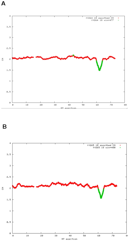
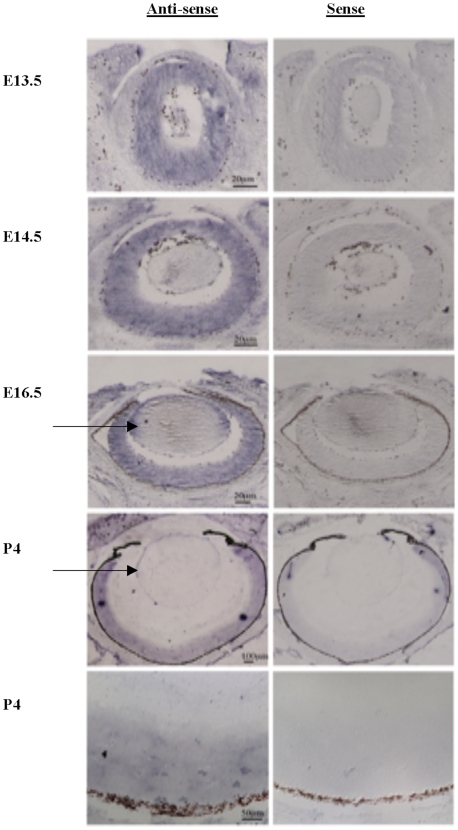
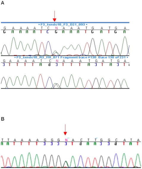
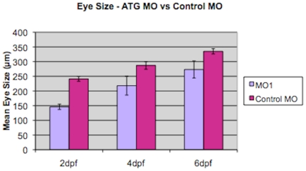
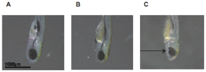
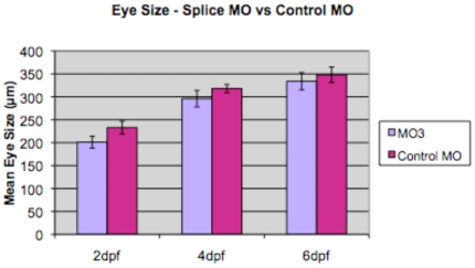
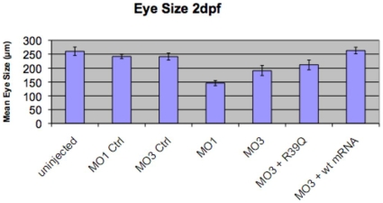
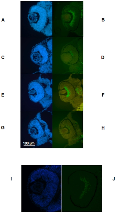
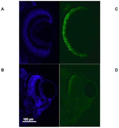
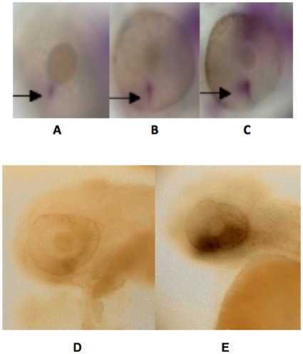
Anophthalmia and microphthalmia are important birth defects, but their pathogenesis remains incompletely understood. We studied a patient with severe unilateral microphthalmia who had a 2.7 Mb deletion at chromosome 18q22.1 that was inherited from his mother. In-situ hybridization showed that one of the deleted genes, TMX3, was expressed in the retinal neuroepithelium and lens epithelium in the developing murine eye. We re-sequenced TMX3 in 162 patients with anophthalmia or microphthalmia, and found two missense substitutions in unrelated patients: c.116G>A, predicting p.Arg39Gln, in a male with unilateral microphthalmia and retinal coloboma, and c.322G>A, predicting p.Asp108Asn, in a female with unilateral microphthalmia and severe micrognathia. We used two antisense morpholinos targeted against the zebrafish TMX3 orthologue, zgc:110025, to examine the effects of reduced gene expression in eye development. We noted that the morphant larvae resulting from both morpholinos had significantly smaller eye sizes and reduced labeling with islet-1 antibody directed against retinal ganglion cells at 2 days post fertilization. Co-injection of human wild type TMX3 mRNA rescued the small eye phenotype obtained with both morpholinos, whereas co-injection of human TMX3(p.Arg39Gln) mutant mRNA, analogous to the mutation in the patient with microphthalmia and coloboma, did not rescue the small eye phenotype. Our results show that haploinsufficiency for TMX3 results in a small eye phenotype and represents a novel genetic cause of microphthalmia and coloboma. Future experiments to determine if other thioredoxins are important in eye morphogenesis and to clarify the mechanism of function of TMX3 in eye development are warranted.


Conflict of interest statement

Competing Interests: The authors have declared that no competing interests exist.

Figures

Figure 1
Figure 1. A 500K Microarray shows a 2.7 Mb deletion of 18q22.1 in the propositus.
Fig. 1A. Graph of smoothed copy number for chromosome 18 from the Affymetrix 500K Array in the propositus, indicating loss of copy number (green color) and demonstrating a chromosome deletion at chromosome 18q22.1. The x-axis shows the nucleotide number from 1–76,117,153 on chromosome 18 and the y-axis shows smoothed copy number. Fig. 1B. Graph of smoothed copy number for chromosome 18 from the Affymetrix 500K Array in the mother of the propositus, indicating a similar loss of copy number (green color) at chromosome 18q22.1. The x-axis shows the nucleotide number from 1–76,117,153 on chromosome 18 and the y-axis shows smoothed copy number.
Figure 2
Figure 2. In-situ hybridization shows strong expression of TMX3 in the developing murine eye.
Fig. 2. In-situ hybridization using antisense and sense riboprobes for TMX3, showing expression in the murine developing eye at E13.5, E14.5, E16.5 and P4. An arrow points to the labeling of the lens epithelium at E16.5 and P4 with the TMX3 antisense probe.
Figure 3
Figure 3. Two patients unrelated to our propositus with microphthalmia have sequence alterations predicting amino acid substitutions in TMX3.
Fig. 3A. Chromatogram showing c.116G>A, predicting p.Arg39Gln in TMX3. Arrow points to the site of the sequence alteration. Fig. 3B. Chromatogram showing c.322G>A, predicting p.Asp108Asn in TMX3. Arrow points to the site of the sequence alteration.
Figure 4
Figure 4. A comparison of eye size in MO1 morpholino-injected larvae and control-injected larvae.
Graph shows mean eye size (measured in µm on y-axis) for MO1 morpholino-injected larvae (MO1; light purple) compared to control-injected larvae (Control MO; dark purple) at three different time periods, 2 dpf, 4 dpf and 6 dpf, labeled on the x-axis. Data are shown as mean +/− one standard deviation, measuring a minimum of 9–20 independent retinas per data point. Analysis using a Two-sample t-test assuming equal variance showed a significant difference at 2 dpf, 4 dpf and 6 dpf (p<0.05 for 2 dpf, 4 dpf and 6 dpf).
Figure 5
Figure 5. Reduced expression of zgc:110025 results in a small eye phenotype.
Fig. 5A. Control injected, wild type zebrafish showing normal eye formation at 6 dpf. Fig. 5B. MO1 morphant zebrafish showing a small eye compared to control at 6 dpf. Fig. 5C. MO1 morphant zebrafish with co-injection of TMX3/(p.Arg39Gln), showing a small ocular coloboma (indicated by arrow) at 6 dpf. Fish are oriented with ventral surface facing left and dorsal surface facing right.
Figure 6
Figure 6. A comparison of eye size in splice morpholino-injected larvae and control-injected larvae.
Graph shows mean eye size (measured in µm on y-axis) for splice morpholino-injected larvae (MO3; light purple) compared to control-injected larvae (Control MO; dark purple) at three different time periods, 2 dpf, 4 dpf and 6 dpf, labeled on the x-axis. Data are shown as mean +/− one standard deviation. A representative single experiment is shown, measuring a minimum of 10–20 independent retinas per data point. Analysis using a Two-sample t-test assuming equal variance showed a significant difference at 2 dpf and 4 dpf (p<0.05 for 2 dpf), but not at 6 dpf (p>0.05).
Figure 7
Figure 7. Co-injection of human wildtype TMX3 mRNA with a splice antisense morpholino rescues morphant eye size.
Data are shown for mean eye size (measured in µm on the y-axis) at 2 dpf for uninjected, control-injected, MO1 and MO3 injected, and MO3 and human wildtype and mutant TMX3 mRNA-injected fish (categories listed on x-axis). A minimum of 10–20 fish were scored for each data point and the data is shown as mean +/− one standard deviation. Rescue of the small eye phenotype can be seen by the similarity in eye measurements obtained for MO3 and human wildtype TMX3 mRNA-injected larvae compared to uninjected and control-injected larvae (p>0.05; two-sample t-test assuming equal variance).
Figure 8
Figure 8. Islet-1 expression is reduced in morphant larvae compared to control larvae at 2 dpf.
Fig. 8A–D. Fig. 8A–B. Labeling of MO1 control-injected zebrafish eye with DAPI (Fig. 8A) and FITC (Fig. 8B) showing labeling for islet-1 at 2 dpf. Fig. 8C–D. Labeling of MO1-injected zebrafish eye with DAPI (Fig. 8C) and FITC (Fig. 8D) showing almost absent labeling for islet-1 at 2 pdf. Fig. 8E–H. Fig. 8E–F. Labeling of MO3 control-injected zebrafish eye with DAPI (Fig. 8E) and FITC (Fig. 8F) showing labeling for islet-1 at 2 pdf. Fig. 8G–H. Labeling of MO3-injected zebrafish eye with DAPI (Fig. 8G) and FITC (Fig. 8H) showing almost absent labeling for islet-1 at 2 pdf, similar to the labeling pattern observed with MO1. Fig. 8I–J. Fig. 8I. MO3-injected zebrafish rescued with human wildtype TMX3 mRNA labeled with DAPI (Fig. 8I) and FITC (Fig. 8J) at 2 dpf, showing labeling with islet-1 antibodies that is similar to labeling in control-injected fish. Ventral is down in all image panels.
Figure 9
Figure 9. Morphant larvae show altered formation of the ventral eye.
Fig. 9A–D. Fig. 9A–B. Staining of control injected zebrafish eye with DAPI and FITC at 6 dpf to image zpr-1 shows a strong signal that can be seen at the ciliary margins of the retina. Fig. 9C–D. Staining of anti-ATG morphant (MO1) zebrafish eye with DAPI and FITC at 6 dpf to image zpr-1 shows absent signal for zpr-1 at the ventral region of the retina in an eye with a coloboma, whereas staining at the dorsal region of the retina appears normal. Fish are oriented so that the ventral surface of the eye is seen inferiorly in each photograph.
Figure 10
Figure 10. Pax2 and Vax2 expression are dysregulated in the region of the choroid fissure in MO1 injected morphant larvae.
Fig. 10. Fig. 10A–C. Representative, uninjected (Fig. 10A), control morpholino injected (Fig. 10B) and MO1 morpholino injected (Fig. 10C) whole embryos hybridized with a Pax2 probe. The region of Pax2 labeling at the site of the choroid fissure (indicated by arrows) is enlarged in the MO1 morphant embryo compared to both the uninjected and control-injected embryos. Fig. 10D. Representative control morpholino injected (Fig. 10D–E) and splice morpholino (MO3) injected (Fig. 10E) whole embryos hybridized with a Vax2 probe. The region of Vax2 labeling (indicated by arrows) is enlarged in the MO3 morphant embryo compared to the control-injected embryo.

References

    1. Martin JA, Kung HC, Mathews TJ, Hoyert DL, Strobino DM, et al. Annual summary of vital statistics: 2006. Pediatrics. 2008;121:788–801. - PubMed
    1. Verma AS, Fitzpatrick DR. Anophthalmia and microphthalmia. Orphanet J Rare Dis. 2007;2:47. - PMC - PubMed
    1. Gregory-Evans CY, Williams MJ, Halford S, Gregory-Evans K. Ocular coloboma: a reassessment in the age of molecular neuroscience. J Med Genet. 2004;41:881–891. - PMC - PubMed
    1. Rainger J, Van Heyningen V, FitzPatrick DR. Development of the Eye. In: Epstein CJ, Erickson RP, Wynshaw-Boris A, editors. Inborn Errors of Development. Oxford: Oxford University Press; 2008. pp. 94–106.
    1. Brown JD, Dutta S, Bharti K, Bonner RF, Munson PJ, et al. Expression profiling during ocular development identifies 2 Nlz genes with a critical role in optic fissure closure. Proc Natl Acad Sci U S A. 2005;106:1462–1467. - PMC - PubMed

Publication types

MeSH terms