. 2010 May 13;6(5):e1000900.
doi: 10.1371/journal.ppat.1000900.

Listeria monocytogenes internalin B activates junctional endocytosis to accelerate intestinal invasion

Affiliations

Listeria monocytogenes internalin B activates junctional endocytosis to accelerate intestinal invasion

Mickey Pentecost et al. PLoS Pathog. .

Abstract

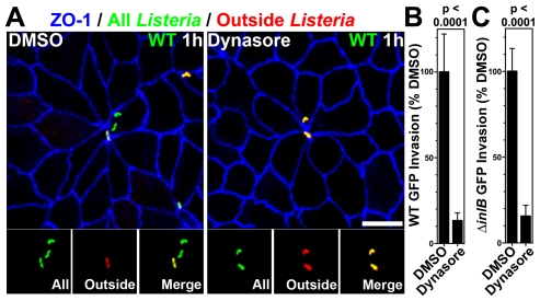
Listeria monocytogenes (Lm) uses InlA to invade the tips of the intestinal villi, a location at which cell extrusion generates a transient defect in epithelial polarity that exposes the receptor for InlA, E-cadherin, on the cell surface. As the dying cell is removed from the epithelium, the surrounding cells reorganize to form a multicellular junction (MCJ) that Lm exploits to find its basolateral receptor and invade. By examining individual infected villi using 3D-confocal imaging, we uncovered a novel role for the second major invasin, InlB, during invasion of the intestine. We infected mice intragastrically with isogenic strains of Lm that express or lack InlB and that have a modified InlA capable of binding murine E-cadherin and found that Lm lacking InlB invade the same number of villi but have decreased numbers of bacteria within each infected villus tip. We studied the mechanism of InlB action at the MCJs of polarized MDCK monolayers and find that InlB does not act as an adhesin, but instead accelerates bacterial internalization after attachment. InlB locally activates its receptor, c-Met, and increases endocytosis of junctional components, including E-cadherin. We show that MCJs are naturally more endocytic than other sites of the apical membrane, that endocytosis and Lm invasion of MCJs depends on functional dynamin, and that c-Met activation by soluble InlB or hepatocyte growth factor (HGF) increases MCJ endocytosis. Also, in vivo, InlB applied through the intestinal lumen increases endocytosis at the villus tips. Our findings demonstrate a two-step mechanism of synergy between Lm's invasins: InlA provides the specificity of Lm adhesion to MCJs at the villus tips and InlB locally activates c-Met to accelerate junctional endocytosis and bacterial invasion of the intestine.


Conflict of interest statement

The authors have declared that no competing interests exist.

Figures

Figure 1
Figure 1. InlB-mediates invasion of intestinal villus tips.
(A) Confocal Z-planes of an ileal villus tip from a mouse infected with 1010 CFU WTm GFP for 4 h and counterstained for F-actin, red, and nuclei, blue. Insets show intracellular Lm with associated F-actin. Depth from the apical cell surface, Z, is indicated. (B) Top panel, a 3D confocal reconstruction of an ileal villus tip of a mouse infected with 1010 CFU WTm GFP for 5 h and counterstained for F-actin, red. Lower panels, Z-planes and insets of intracellular Lm with associated F-actin. (C) Top panel, a 3D confocal reconstruction of an ileal villus tip of a mouse infected with 1010 CFU ΔinlB m GFP for 5 h and counterstained for F-actin, red. Lower panels, Z-planes and insets of intracellular Lm associated with F-actin. (D–E) Coinfection with 5×10⁹ CFU each WTm GFP and ΔinlB m Lm for 6 h. Tissue was stained with phalloidin for F-actin, blue, and for all Lm, red. (D) 3D confocal reconstruction of infected villus tips. (E) Quantification of Listeria per infected villus tip for 3 mice. (F–H) Coinfection with 5×10⁹ CFU each WTm and ΔinlB m GFP Lm for 6 h. Tissue was stained with phalloidin for F-actin, blue, and for all Listeria, red. (F) Top left, 3D confocal reconstruction of infected villus tips. Top right, F-actin staining from the top left panel is omitted to show all Lm. Bottom panels, zoomed insets from top right. (G) A rare villus tip infected with both WTm and ΔinlB m GFP Lm. (H) Quantification of Listeria per infected villus tip. Scale bars, 10 µm.
Figure 2
Figure 2. InlB and c-Met accelerate invasion of multicellular junctions.
(A) Confluent MDCK monolayers were infected with WT or ΔinlB Lm at an MOI of 100. Adhesion after 10 min of infection determined by quantification of all cell-associated CFUs, left, or invasion determined by quantification of viable CFUs of intracellular bacteria after gentamicin treatment, right. Mean and SD from a representative experiment performed in triplicate is shown. (B) Polarized MDCK monolayers were infected with an MOI of 10 of GFP-expressing Lm. Invasion at multicellular junctions was visualized with anti-ZO-1 antibodies, blue. To evaluate intracellular versus extracellular bacteria, extracellular adherent GFP-expressing Lm were stained before permeabilization in red. External Lm thus appear as a combination of red/green or yellow. Scale bars 10 µm. (C) Quantification of intracellular bacteria from monolayers as in B. Mean and SD from three 60X fields are shown. (D) Polarized MDCK monolayers infected with an MOI of 10 were fixed at the indicated time points post infection and the number of Listeria per plaque quantified by confocal immunofluorescence analysis. Exponential curves were fit to all data points per strain. WT Td = 1.25 h; R2 = 0.818. ΔinlB Td = 1.26 h; R2 = 0.898. (E) Confluent MDCK monolayers were treated with either DMSO or a c-Met inhibitor prior to infection with WT or ΔinlB Lm at an MOI 100 and quantification of viable CFUs of intracellular bacteria after gentamicin treatment. Mean and SD from triplicate samples are shown. (F) To determine whether the invasion defect of ΔinlB could be rescued, confluent MDCK monolayers were either untreated or treated with c-Met inhibitor prior and during infection with a 1∶1 ratio of WT∶WT, as a control, or WT∶ΔinlB at an MOI of 100. The ratio of the strains, competitive index (C.I.), recovered after gentamicin treatment was determined.
Figure 3
Figure 3. InlB and HGF accelerate dynamin-dependent endocytosis at multicellular junctions.
(A) Extended focus views of polarized MDCK monolayers treated with DMSO 30 minutes prior to and including apical treatment with InlB or HGF for 1 h, then apical treatment with dextran, a fluid phase internalization marker, red, for 30 minutes. Monolayers were counterstained for ZO-1, green. (B) Quantification of dextran fluorescence in 50 µm×50 µm regions centered at multicellular junctions (MCJ) or at regions without multicellular junctions (Non-MCJ) of DMSO treated monolayers. (C) Quantification of dextran puncta in 50 µm×50 µm regions centered at MCJs in DMSO treated monolayers. (D) Quantification of dextran fluorescence in all puncta analyzed at MCJs. (E) Single confocal Z-plane through polarized E-cadherin-GFP MDCK cells treated apically with dextran, red, for 30 minutes and counterstained for ZO-1, blue. (F) 3D rendered confocal images E-cadherin-GFP MDCK cells treated apically with InlB or HGF and with dextran, red, for 30 minutes. (G) Extended focus views of polarized MDCK cells treated with dynasore, to inhibit dynamin function, 30 minutes prior to and including apical treatment with InlB or HGF for 1 h, then apical treatment with dextran, red, for 30 minutes. Monlayers were counterstained for ZO-1, green. (H) Quantification of dextran fluorescence in 50 µm×50 µm regions centered at multicellular junctions (MCJ) or at regions without multicellular junctions (Non-MCJ) of dynasore treated monolayers; data normalized to B and displayed with a different scale. Scale bars 10 µm.
Figure 4
Figure 4. Dynamin-dependent invasion of MCJs.
(A) Polarized MDCK cells were pretreated with DMSO or dynasore for 30 minutes and then infected with GFP-expressing WT Lm for 1 h. To evaluate intracellular versus extracellular bacteria, we performed an outside staining where extracellular adherent GFP-expressing Lm were stained before permeabilization in red. Intracellular Lm are green and external Lm are a combination of red/green or yellow. Monolayers were counterstained with antibodies to ZO-1, blue. Scale bars 10 µm. (B) Quantification of relative WT Lm invasion of Polarized MDCK monolayers pretreated with DMSO or dynasore for 1 h. Relative mean and SD of intracellular CFUs recovered after gentamicin treatment are shown. p<0.0001. (C) Quantification of relative ΔinlB Lm invasion of Polarized MDCK monolayers treated with DMSO or dynasore. Relative mean and SD of intracellular CFUs recovered after gentamicin treatment are shown. p<0.0001.
Figure 5
Figure 5. InlB enhances endocytosis at the intestinal villus tips.
Fluorescent dextran, a fluid phase internalization marker, with or without InlB was incubated for 45 minutes in mouse ileal loops. (A) 3D confocal reconstructions of villus tips from an ileal loop incubated with dextran, red. Tissue was counterstained with phalloidin for F-actin, green. (B) Higher magnification villus tip from an ileal loop incubated with dextran, and a single Z-plane from inset showing puncta of intracellular dextran. (C) Single Z-plane from confocal image of villus tips from a loop incubated with dextran, red, and counterstained with anti-E-cadherin antibodies, green, and with phalloidin for F-actin, blue. Inset shows higher magnification of dextran puncta and intracellular E-cadherin. (D) 3D confocal reconstructions of villus tips from an ileal loop incubated with dextran, red, and InlB. Tissue was counterstained with phalloidin for F-actin, green. (E) Higher magnification villus tip from an ileal loop incubated with dextran, red, and InlB, and a single Z-plane from inset showing puncta of intracellular dextran. (F) Single Z-plane from confocal image of villus tips from a loop incubated with dextran, red, and InlB and counterstained with anti-E-cadherin antibodies, green, and phalloidin for F-actin, blue. Inset shows higher magnification of three separate dextran puncta and intracellular E-cadherin. Scale bars 10 µm. (G) Quantification of dextran fluorescence per villus tip from ileal loops incubated with dextran only (−) or dextran and InlB (+). (H) Quantification of dextran puncta per villus tip from ileal loops incubated with dextran only (−) or dextran and InlB (+). (I) Quantification of dextran fluorescence from all puncta analyzed from villus tips of ileal loops incubated with dextran only (−) or dextran and InlB (+).
Figure 6
Figure 6. Diagram of Lm invasion coupled to junctional remodeling by endocytosis at the MCJ.
(A) Lm find exposed E-cadherin at multicellular junctions (MCJs) when extruded cells are displaced from the epithelium. New tight junctions (TJs) and adherens junctions (AJs) are formed below the extruded cell at the invagination of MCJs. The old junctions and intercellular-adhesions are removed by endocytosis. RTKs, receptor tyrosine kinases. (B) Close up diagram of the region of bacterial invasion at the MCJ. Left: after binding to E-cadherin via InlA, InlB activates c-Met which is also abnormally exposed to the apical surface at MCJs. Activation of c-Met locally by InlB at the MCJ increases the rate of endocytosis of Lm. Right: endocytic removal of the old junctions parallels Lm uptake. Experimentally, c-Met activation by HGF may increase the rate of formation and the size of endosomes. E-cadherin, yellow; InlA, magenta; c-Met, blue; InlB, orange; HGF, white; dynamin, purple; a simplified tight junction, red.

Similar articles

Cited by

References

    1. Vazquez-Boland JA, Kuhn M, Berche P, Chakraborty T, Dominguez-Bernal G, et al. Listeria pathogenesis and molecular virulence determinants. Clin Microbiol Rev. 2001;14:584–640. - PMC - PubMed
    1. Dramsi S, Biswas I, Maguin E, Braun L, Mastroeni P, et al. Entry of Listeria monocytogenes into hepatocytes requires expression of inIB, a surface protein of the internalin multigene family. Mol Microbiol. 1995;16:251–261. - PubMed
    1. Gaillard JL, Berche P, Frehel C, Gouin E, Cossart P. Entry of L. monocytogenes into cells is mediated by internalin, a repeat protein reminiscent of surface antigens from gram-positive cocci. Cell. 1991;65:1127–1141. - PubMed
    1. Temm-Grove CJ, Jockusch BM, Rohde M, Niebuhr K, Chakraborty T, et al. Exploitation of microfilament proteins by Listeria monocytogenes: microvillus-like composition of the comet tails and vectorial spreading in polarized epithelial sheets. J Cell Sci. 1994;107 (Pt 10):2951–2960. - PubMed
    1. Ray K, Marteyn B, Sansonetti PJ, Tang CM. Life on the inside: the intracellular lifestyle of cytosolic bacteria. Nat Rev Microbiol. 2009;7:333–340. - PubMed

Publication types

MeSH terms