. 2010 May 13;6(5):e1000948.
doi: 10.1371/journal.pgen.1000948.

Defective resection at DNA double-strand breaks leads to de novo telomere formation and enhances gene targeting

Affiliations

Defective resection at DNA double-strand breaks leads to de novo telomere formation and enhances gene targeting

Woo-Hyun Chung et al. PLoS Genet. .

Abstract

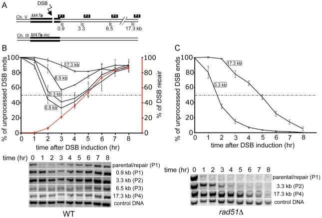
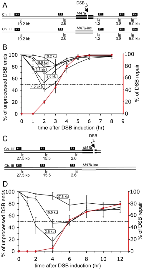
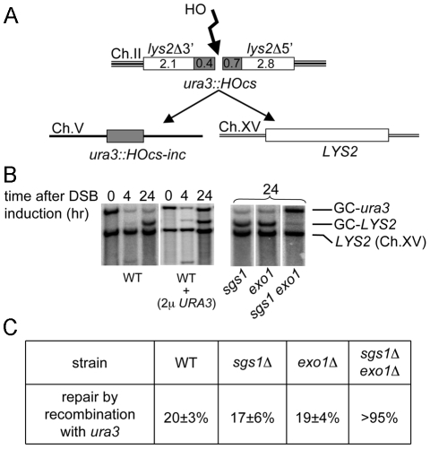
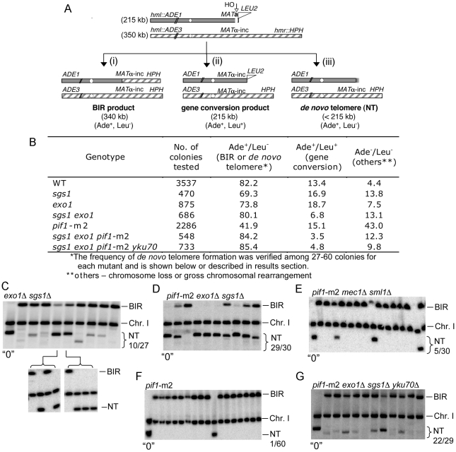
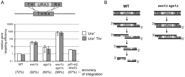
The formation of single-stranded DNA (ssDNA) at double-strand break (DSB) ends is essential in repair by homologous recombination and is mediated by DNA helicases and nucleases. Here we estimated the length of ssDNA generated during DSB repair and analyzed the consequences of elimination of processive resection pathways mediated by Sgs1 helicase and Exo1 nuclease on DSB repair fidelity. In wild-type cells during allelic gene conversion, an average of 2-4 kb of ssDNA accumulates at each side of the break. Longer ssDNA is formed during ectopic recombination or break-induced replication (BIR), reflecting much slower repair kinetics. This relatively extensive resection may help determine sequences involved in homology search and prevent recombination within short DNA repeats next to the break. In sgs1Delta exo1Delta mutants that form only very short ssDNA, allelic gene conversion decreases 5-fold and DSBs are repaired by BIR or de novo telomere formation resulting in loss of heterozygosity. The absence of the telomerase inhibitor, PIF1, increases de novo telomere pathway usage to about 50%. Accumulation of Cdc13, a protein recruiting telomerase, at the break site increases in sgs1Delta exo1Delta, and the requirement of the Ku complex for new telomere formation is partially bypassed. In contrast to this decreased and alternative DSB repair, the efficiency and accuracy of gene targeting increases dramatically in sgs1Delta exo1Delta cells, suggesting that transformed DNA is very stable in these mutants. Altogether these data establish a new role for processive resection in the fidelity of DSB repair.


Conflict of interest statement

The authors have declared that no competing interests exist.

Figures

Figure 1
Figure 1. Measurement of resection in DSB–induced ectopic recombination.
(A) Schematic representation of the ectopic recombination assay between MAT a on chromosome V and MAT a-inc on chromosome III. Positions of EcoRI sites (E) and DNA probes used for hybridization with respect to the HO recognition site on chromosome V are shown (tGI354). MAT a and MAT a-inc share 1.9 kb of homology in total. (B) Plot showing the percentage of unprocessed 5′ strand for each EcoRI site and the corresponding Southern blots are shown. Kinetics of DSB repair product formation is indicated by the red line. Plotted values are the mean values ±SD from three independent experiments. (C) 5′ strand processing was monitored and plotted from an ectopic recombination assay in rad51Δ cells (tGI379) as in (B).
Figure 2
Figure 2. Kinetics of repair determine the amount of ssDNA during DSB repair.
(A) Schematic representation of the allelic recombination assay between MAT a and MATα-inc loci on chromosome III. Positions of EcoRI (E) and HindIII (H) sites and DNA probes used for Southern hybridization to analyze 5′ strand processing with respect to the HO recognition site are shown (yGI234). (B) Plot showing the percentage of unprocessed 5′ strand for each restriction site. Plotted values are the mean values ±SD from three independent experiments. Kinetics of DSB repair is indicated by the red line. (C) Schematic diagram of the BIR assay used for resection analysis. (D) 5′ strand processing was analyzed in the BIR repair strain (AM1003) as described in (B).
Figure 3
Figure 3. Resection at DSB ends determines the choice of recombination donor.
(A) Schematic diagram of the competition assay used to determine whether sequences immediately next to the break (ura3) or sequences farther from the break (lys2) were used during gene conversion . Details of the assay are described in the text. Sequence sizes are in kilobase pairs. (B) Southern blotting analysis of the competition gene conversion assay shown in (A). The LYS2 gene was used as a probe. The positions of two gene conversion products called GC-ura3 and GC-LYS2 are indicated. (C) Frequencies of the choice of the ura3::HOcs-inc donor for DSB repair measured at 24 hr timepoint as the density of the band corresponding to the GC-ura3 product divided by the combined density of both GC-ura3 and GC-LYS2 products in different mutants are shown in the table.
Figure 4
Figure 4. De novo telomeres are frequently formed at poorly processed DSB ends.
(A) Schematic diagram showing chromosome structure of the disomic strain (AM1003) that carries an original chromosome III (350 kb) and a fragmented chromosome III (215 kb). Sequences distal to the HO recognition site were replaced by the LEU2 gene and telomere sequences as described . Three major pathways of repair with their respective product sizes are shown: BIR, gene conversion and de novo telomere addition (NT). Products can be distinguished by markers and/or by sizes on PFGE gels. (B) Frequency of DSB repair by BIR and gene conversion in the AM1003 strain and its derivatives is shown in the table. (C) PFGE analysis of products from Ade+ Leu colonies exo1Δ sgs1Δ, (D) pif1-m2 exo1Δ sgs1Δ, (E) pif1-m2 med1Δ sml1Δ (F) pif1-m2 and (G) pif1-m2 exo1Δ sgs1Δ yku70Δ mutant strains. To separate BIR repair products and telomere-added products (NT) in the exo1Δ sgs1Δ strain (C) originating from one survivor we streaked single colonies on YEPD plates and analyzed products from individual colonies by subsequent PFGE. Genomic DNA of products repaired by de novo telomere addition was purified and used for sequencing to identify telomere addition sites [(C,D) and Figure 5]. “0” indicates control with no break induction. Position of chromosome I (Chr.I) that carries ade1–1 allele and is detected with ADE1 probe is shown.
Figure 5
Figure 5. Analysis of telomere formation in exo1Δ sgs1Δ mutants.
(A) The positions of new telomere addition in 14 products from exo1Δ sgs1Δ (5 triangles) and pif1-m2 exo1Δ sgs1Δ (9 circles) cells were determined by sequencing. (B) Exact position and sequence where telomeres were added is presented in the table. (C) ChIP analysis of Cdc13-13Myc binding to the region flanking the DSB was conducted in wild-type, pif1-m2, exo1Δ sgs1Δ and pif1-m2 exo1Δ sgs1Δ strains using polyclonal anti-myc antiserum. Samples were collected before and 8 h after DSB induction. Immunoprecipitated DNAs were amplified by qPCR using primer pairs to amplify a region located about 1 kb away from the HO break site. The 8 h time point IP values were normalized to the time 0 samples to yield the fold-IP values plotted on the Y-axis for the wild-type and each mutant strain. Error bars represent one standard error of the mean for three independent experiments.
Figure 6
Figure 6. Elimination of enzymes that degrade DNA ends greatly stimulates gene targeting.
(A) A 2.1 kb thr4::URA3 cassette was used for a gene replacement assay in wild-type and mutant strains lacking one or more enzymes involved in DSB resection. Efficiency and accuracy of gene targeting measured as the amount of Ura+ or Ura+ Thr colonies is displayed. Error bars represent standard deviation for three independent experiments. (B) Hypothetical model presenting gene targeting in cells exhibiting normal processing of DNA ends and in cells with decreased resection. Extensive resection exposes the nonhomologous URA3 marker sequence and eventually degrades the transformed cassette. Exposed 3′ homologous tails are likely to be degraded faster in wild-type cells than in sgs1Δ exo1Δ mutant cells.

References

    1. San Filippo J, Sung P, Klein H. Mechanism of eukaryotic homologous recombination. Annu Rev Biochem. 2008;77:229–257. - PubMed
    1. Symington LS. Role of RAD52 epistasis group genes in homologous recombination and double-strand break repair. Microbiol Mol Biol Rev. 2002;66:630–670. - PMC - PubMed
    1. Gravel S, Chapman JR, Magill C, Jackson SP. DNA helicases Sgs1 and BLM promote DNA double-strand break resection. Genes Dev. 2008;22:2767–2772. - PMC - PubMed
    1. Mimitou EP, Symington LS. Sae2, Exo1 and Sgs1 collaborate in DNA double-strand break processing. Nature. 2008;455:770–774. - PMC - PubMed
    1. Zhu Z, Chung WH, Shim EY, Lee SE, Ira G. Sgs1 helicase and two nucleases dna2 and exo1 resect DNA double-strand break ends. Cell. 2008;134:981–994. - PMC - PubMed

Publication types

Substances