. 2010 May 13;6(5):e1000781.
doi: 10.1371/journal.pcbi.1000781.

Impact of dendritic size and dendritic topology on burst firing in pyramidal cells

Affiliations

Impact of dendritic size and dendritic topology on burst firing in pyramidal cells

Ronald A J van Elburg et al. PLoS Comput Biol. .

Abstract

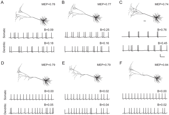
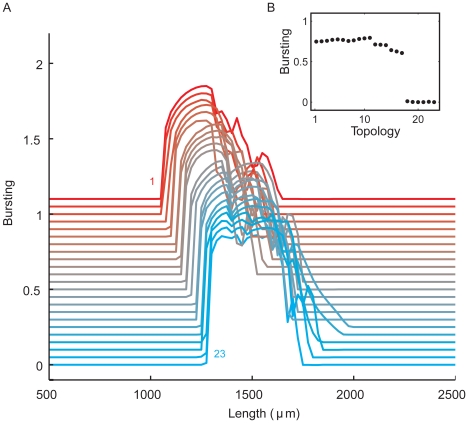
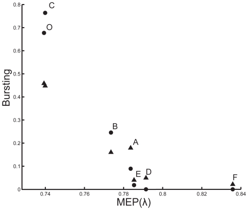
Neurons display a wide range of intrinsic firing patterns. A particularly relevant pattern for neuronal signaling and synaptic plasticity is burst firing, the generation of clusters of action potentials with short interspike intervals. Besides ion-channel composition, dendritic morphology appears to be an important factor modulating firing pattern. However, the underlying mechanisms are poorly understood, and the impact of morphology on burst firing remains insufficiently known. Dendritic morphology is not fixed but can undergo significant changes in many pathological conditions. Using computational models of neocortical pyramidal cells, we here show that not only the total length of the apical dendrite but also the topological structure of its branching pattern markedly influences inter- and intraburst spike intervals and even determines whether or not a cell exhibits burst firing. We found that there is only a range of dendritic sizes that supports burst firing, and that this range is modulated by dendritic topology. Either reducing or enlarging the dendritic tree, or merely modifying its topological structure without changing total dendritic length, can transform a cell's firing pattern from bursting to tonic firing. Interestingly, the results are largely independent of whether the cells are stimulated by current injection at the soma or by synapses distributed over the dendritic tree. By means of a novel measure called mean electrotonic path length, we show that the influence of dendritic morphology on burst firing is attributable to the effect both dendritic size and dendritic topology have, not on somatic input conductance, but on the average spatial extent of the dendritic tree and the spatiotemporal dynamics of the dendritic membrane potential. Our results suggest that alterations in size or topology of pyramidal cell morphology, such as observed in Alzheimer's disease, mental retardation, epilepsy, and chronic stress, could change neuronal burst firing and thus ultimately affect information processing and cognition.


Conflict of interest statement

The authors have declared that no competing interests exist.

Figures

Figure 1
Figure 1. Pyramidal cell and burst firing.
A, Reconstructed layer 5 pyramidal cell from cat visual cortex (adapted from [18]). Scale bar: 100 µm. The apical tree has a total length of 36865 µm, a total surface area of 25828 µm2, a total volume of 13292 µm3, a root segment with a diameter of 8.5 µm, and 41 terminal segments with diameters in the range 0.30–1.33 µm. The 10 basal dendrites have a total length of 10232 µm, a total surface area of 27396 µm2, a total volume of 7650 µm3, root segments with diameters in the range 1–4 µm, and in total 35 terminal segments with diameters in the range 0.59–1.31 µm. The MEP value (in units of the electrotonic length constant) of the apical dendrite is 0.74. The input conductance of the cell without basal dendrites is 7.9 µS and with basal dendrites 19.9 µS. B, Firing pattern (voltage trace) evoked in the cell of A by a continuous current injection at the soma (somatic stimulation) or by random activation of excitatory synapses distributed across the apical dendritic tree (dendritic stimulation). The burst measure B quantifies the degree of bursting. Scale bar: 100 ms, 50 mV. C, Examples of spike patterns illustrating different values of B. From top to bottom: a perfectly regular pattern without bursts, a random (Poisson) pattern, and two different burst patterns. The B value of both the regular and the Poisson spike train is zero, indicating complete lack of bursting. The two spike patterns with bursting illustrate that the higher the ratio of inter- to intraburst interspike intervals, the higher the value of B.
Figure 2
Figure 2. Morphologically simplified cells.
A, The set of 23 neurons consisting of all the topologically different trees with 8 terminal segments. The neurons are ordered according to the degree of symmetry of their branching structure (see Methods), with 1 having the most asymmetric and 23 the most symmetric tree. B, In the description of the morphology of a dendritic tree, we distinguish between terminal or end segments (which terminate in a tip) and intermediate segments (which terminate in a branch point). The root segment is the intermediate segment that is connected to the soma. The connectivity pattern of the segments is called the topology of a tree. At the right is the description of this tree in the notation we employed to order the trees.
Figure 3
Figure 3. Both with somatic and with dendritic stimulation, pyramidal cell burst firing decreases as the apical dendrite becomes shorter.
A, The degree of bursting, as measured by the burst measure B, against the total length of the apical dendrite. Starting with the cell in Fig. 1A, we reduced the total length by successively pruning terminal segments off the apical dendrite. B, Examples from the experiment in A showing that the removal of only a few small terminal segments from the apical dendritic tree can change the firing state of the cell. Morphology of pruned pyramidal cells, and voltage traces for both somatic and dendritic stimulation. Left, Bursting cells (Top, 9772 µm; Bottom, 8925 µm). Right, Non-bursting cells (Top, 8184 µm; Bottom, 6927 µm). Scale bar: 100 ms, 50 mV. Scale bar (anatomy): 100 µm. Arrows in the bursting cells indicate the branches that are shorter or absent in the non-bursting cells.
Figure 4
Figure 4. Both with somatic and with dendritic stimulation, pyramidal cell burst firing disappears when the apical dendrite becomes either too large or too small.
Using the cell from Fig. 1A, we varied the size of the apical dendrite by scaling the entire apical dendrite, thus keeping the dendritic arborization intact. A, The degree of bursting against the factor by which the length of all the apical dendritic segments was multiplied. B, Voltage traces obtained for different sizes of the apical dendrite. Left, somatic stimulation. Right, dendritic stimulation. Scale bars: 100 ms, 50 mV.
Figure 5
Figure 5. Both with somatic and with dendritic stimulation, dendritic topology affects pyramidal cell burst firing.
Using the cell from Fig. 1A, we varied the topology of the apical dendritic tree by swapping branches within the tree. Thus, all the pyramidal cells shown have exactly the same total dendritic length and dendritic surface area and differ only in the topology of their apical dendrite (basal dendrites are the same). Voltage traces for three bursting cells (A–C), and three non-bursting cells (D–F). Scale bar: 100 ms, 50 mV. Scale bar (anatomy): 100 µm. MEP values indicate the mean electrotonic path length of the apical dendritic tree.
Figure 6
Figure 6. Dendritic topology influences burst firing, as demonstrated in the set of morphologically simplified neurons (Fig. 2) using somatic stimulation.
The segment diameters in the trees obey Rall's power law. A, Examples from the set of 23 tree topologies together with corresponding voltage traces. Each tree has a total dendritic length of 1750 µm. Scale bar: 100 ms, 100 mV. B, The range of tree sizes where burst firing occurs is modulated by dendritic topology. For all the 23 tree topologies, the degree of bursting is shown as a function of the total length of the dendritic tree, plotted with an offset (for all horizontal lines B = 0) for clarity. The topologies are ordered as in Fig. 2, with the top line representing topology 1, and the bottom line topology 23. The total dendritic length of a given tree topology was varied by multiplying the lengths of all its segments by the same factor. C, Bursting as a function of topology at a total dendritic length of 1750 µm. D, For topology 1, voltage traces at different total dendritic lengths, together with B values indicating the degree of bursting. From top to bottom: 1000, 1300, 1600, 1900, 2200, 2500, and 2800 µm. Scale bar: 100 ms, 100 mV.
Figure 7
Figure 7. Dendritic topology influences burst firing, as demonstrated in the set of morphologically simplified neurons using dendritic stimulation.
The segment diameters in the trees obey Rall's power law. A, Examples from the set of 23 tree topologies together with corresponding voltage traces. Each tree has a total dendritic length of 1900 µm. Scale bar: 100 ms, 100 mV. B, As with somatic stimulation, the range of tree sizes where burst firing occurs is affected by dendritic topology. For all the 23 tree topologies, the degree of bursting is shown as a function of the total length of the dendritic tree, plotted with an offset for clarity. The topologies are ordered as in Fig. 2, with the top line representing topology 1, and the bottom line topology 23. The total dendritic length of a given tree topology was varied by multiplying the lengths of all its segments by the same factor. C, Bursting as a function of topology at a total dendritic length of 1900 µm. D, For topology 1, voltage traces at different total dendritic lengths, together with B values indicating the degree of bursting. From top to bottom: 1000, 1400, 1800, 2200, 2600, 3000, and 3400 µm. Scale bar: 100 ms, 100 mV.
Figure 8
Figure 8. Dendritic topology influences burst firing also if all segment diameters are uniform throughout the whole tree.
Thus, all the 23 tree topologies have the same total dendritic surface area as well the same total dendritic length. A, For all the 23 tree topologies, the degree of bursting is shown as a function of the total length of the dendritic tree, plotted with an offset for clarity, and using somatic stimulation. The topologies are ordered as in Fig. 2, with the top line representing topology 1, and the bottom line topology 23. The total dendritic length of a given tree topology was varied by multiplying the lengths of all its segments by the same factor. B, Bursting as a function of topology at a total dendritic length of 1250 µm.
Figure 9
Figure 9. Mean electrotonic path length, but not input conductance, correlates with the region of burst firing for trees with uniform segment diameters and under somatic stimulation.
Except for the white contour lines, the left and right panels are identical and show the degree of burst firing (color coded) as a function of both dendritic topology and total dendritic length. In the left panel, contour lines of equal input conductance (IC, in µS) are superimposed. These contour lines show all the combinations of dendritic length and dendritic topology that result in a given input conductance. Similarly, in the right panel, contour lines of equal mean electrotonic path length (MEP, in units of the electrotonic length constant) are superimposed. These contour lines show all the combinations of dendritic length and dendritic topology that result in a given MEP value. The onset and cessation of bursting are strongly associated with mean electrotonic path length, but not with input conductance.
Figure 10
Figure 10. Mean electrotonic path length correlates with the region of burst firing for trees whose segment diameters obey Rall's power law.
The degree of burst firing (color coded) is shown as a function of both dendritic topology and total dendritic length, under somatic stimulation (Left) and dendritic stimulation (Right). The superimposed white lines are contour lines of equal mean electrotonic path length (MEP, in units of the electrotonic length constant), showing all the combinations of dendritic length and dendritic topology that result in a given MEP value. Especially the onset of burst firing is strongly associated with mean electrotonic path length.
Figure 11
Figure 11. The importance of electrotonic distance for burst firing and the impact of dendritic topology illustrated with a fully asymmetrical and a fully symmetrical tree.
A, B, At this dendritic size, the asymmetrical tree (A) generates bursts, whereas the symmetrical tree (B) produces single spikes. The segment diameters in the trees obey Rall's power law, and both trees have the same total dendritic length (1600 µm). Scale bars: 100 ms, 20 mV. The ticks on top of the action potentials in A and B indicate the spikes that are shown at t+0 and t+13 in panel C. C, The membrane potential evolution over time in the asymmetrical tree (top row) and the symmetrical tree (bottom row) along the dendritic paths indicated in A and B. Time is relative to the first spike (at 0 ms), and membrane position on the x-axis runs from soma to the tip of the terminal segment. Because the distance between soma and terminal segment is smaller in the symmetrical than in the asymmetrical tree, the membrane potential evolution in the symmetrical tree has less spatial differentiation, the membrane potential reaches a lower value at the distal end, and the distal membrane potential start decreasing earlier in time, so that the return current from dendrites to soma reaches the soma at a time when the delayed-rectifier K+ channels are still open, preventing the generation of a second spike. See further Results.
Figure 12
Figure 12. Burst firing in the pyramidal cells of Fig. 5 correlates strongly with mean electrotonic path length (MEP, in units of the electrotonic length constant), both with somatic stimulation (circles; r = −0.97) and dendritic stimulation (triangles; r  = −0.99).
(In the calculation of the correlation, we excluded the two MEP values around 0.84, thus ignoring the part of the curve that is clearly flat). The letters A–F correspond to those in Fig. 5, and O indicates the unperturbed pyramidal cell of Fig. 1.

References

    1. Llinás RR. The intrinsic electrophysiological properties of mammalian neurons: insights into central nervous system function. Science. 1988;242:1654–1664. - PubMed
    1. Connors BW, Gutnick MJ. Intrinsic firing patterns of diverse neocortical neurons. Trends Neurosci. 1990;13:99–104. - PubMed
    1. Contreras D. Electrophysiological classes of neocortical neurons. Neural Networks. 2004;17:633–646. - PubMed
    1. Krahe R, Gabbiani F. Burst firing in sensory systems. Nature Reviews Neuroscience. 2004;5:13–23. - PubMed
    1. Eggermont JJ, Smith GM. Burst-firing sharpens frequency-tuning in primary auditory cortex. Neuroreport. 1996;7:753–757. - PubMed

Publication types

LinkOut - more resources