

Arteriovenous malformation causing ileocecal variceal bleeding in liver cirrhosis: case report and review of the literature
- PMID: 20485612
- PMCID: PMC2871570
- DOI: 10.5009/gnl.2008.2.1.54
Arteriovenous malformation causing ileocecal variceal bleeding in liver cirrhosis: case report and review of the literature
Abstract
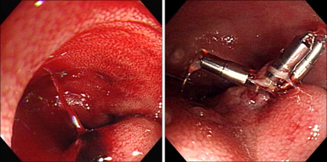
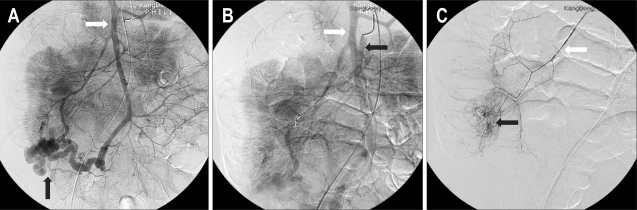
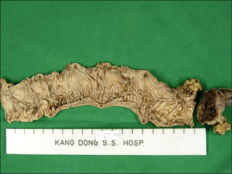
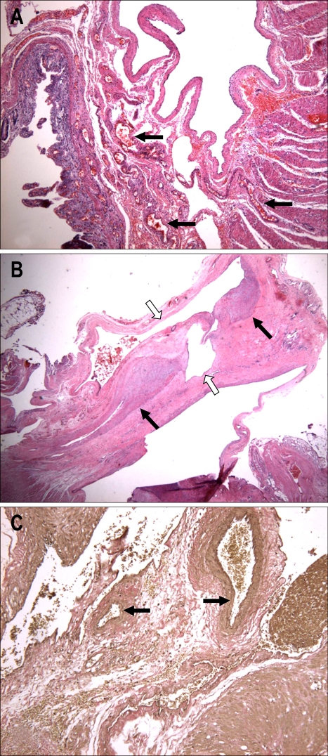
Varices that occur at sites other than the esophagogastric area are termed ectopic varices. An ileal varix is a very rare cause of lower gastrointestinal bleeding. Although ileal varices are generally associated with prior intra-abdominal surgery and adhesions, an arteriovenous malformation (AVM) in the ileocecal area can cause ileal varices and bleeding in patients with portal hypertension who have not received previous intra-abdominal surgery, which is due to an intestinal or colonic AVM dilating the collateral veins and further aggravating portal hypertension. Surgical treatment should be considered in patients with massive ectopic variceal bleeding. We report a case of massive ileocecal variceal bleeding associated with an AVM that occurred in a patient with alcoholic liver cirrhosis.
Keywords: Arteriovenous malformation; Ileocecal varix; Portal hypertension.
Figures
Similar articles
-
Ileocecal Mesentery Arteriovenous Malformation as a Rare Cause of Ectopic Variceal Bleeding in a 58-Year-Old Male With Cirrhosis.Cureus. 2023 Sep 22;15(9):e45785. doi: 10.7759/cureus.45785. eCollection 2023 Sep. Cureus. 2023. PMID: 37872923 Free PMC article.
-
Ectopic gastrointestinal variceal bleeding with portal hypertension.World J Gastrointest Surg. 2017 Dec 27;9(12):288-292. doi: 10.4240/wjgs.v9.i12.288. World J Gastrointest Surg. 2017. PMID: 29359035 Free PMC article.
-
Ectopic variceal bleeding due to portosystemic shunt via dilated mesenteric veins and a varicous left ovarian vein : case report and literature review of ectopic varices.Acta Gastroenterol Belg. 2017 Jul-Sep;80(3):388-395. Acta Gastroenterol Belg. 2017. PMID: 29560669 Review.
-
Bleeding esophageal varices associated with pancreatic arteriovenous malformation.World J Surg. 1991 Jan-Feb;15(1):57-60; discussion 60-1. doi: 10.1007/BF01658962. World J Surg. 1991. PMID: 1994606
-
Natural history of portal hypertension in patients with cirrhosis.Clin Liver Dis. 2001 Aug;5(3):645-63. doi: 10.1016/s1089-3261(05)70186-0. Clin Liver Dis. 2001. PMID: 11565135 Review.
Cited by
-
Proctalgia secondary to rectal arteriovenous malformation and inferior mesenteric vein stenosis in a patient post liver transplant.CVIR Endovasc. 2021 Jan 5;4(1):5. doi: 10.1186/s42155-020-00196-1. CVIR Endovasc. 2021. PMID: 33400018 Free PMC article.
-
Laparoscopic sigmoid colectomy for transverse colonic varices due to an inferior mesenteric arteriovenous fistula.Surg Case Rep. 2024 May 3;10(1):112. doi: 10.1186/s40792-024-01911-z. Surg Case Rep. 2024. PMID: 38700649 Free PMC article.
-
Multiple De Novo Cerebral Arteriovenous Malformations in a Patient with Alcoholic Liver Cirrhosis.J Belg Soc Radiol. 2022 May 27;106(1):53. doi: 10.5334/jbsr.2822. eCollection 2022. J Belg Soc Radiol. 2022. PMID: 35651917 Free PMC article.
-
Ileocecal Mesentery Arteriovenous Malformation as a Rare Cause of Ectopic Variceal Bleeding in a 58-Year-Old Male With Cirrhosis.Cureus. 2023 Sep 22;15(9):e45785. doi: 10.7759/cureus.45785. eCollection 2023 Sep. Cureus. 2023. PMID: 37872923 Free PMC article.
-
Atypical Polyps Presenting With Occult Bleeding.ACG Case Rep J. 2019 Mar 6;6(2):e00017. doi: 10.14309/crj.0000000000000017. eCollection 2019 Feb. ACG Case Rep J. 2019. PMID: 31616719 Free PMC article. No abstract available.
References
-
- Lewis P, Warren BF, Bartolo DC. Massive gastrointestinal haemorrhage due to ileal varices. Br J Surg. 1990;77:1277–1278. - PubMed
-
- Kotfila R, Trudeau W. Extraesophageal varices. Dig Dis. 1998;16:232–241. - PubMed
-
- Vescia FG, Babb RR. Colonic varices: a rare, but important cause of gastrointestinal hemorrhage. J Clin Gastroenterol. 1985;7:63–65. - PubMed
-
- Ohtani T, Kajiwara E, Suzuki N, et al. Ileal varices associated with recurrent bleeding in a patient with liver cirrhosis. J Gastroenterol. 1999;34:264–268. - PubMed
-
- Ueda J, Yoshida H, Mamada Y, et al. Successful emergency enterectomy for bleeding ileal varices in a patient with liver cirrhosis. J Nippon Med Sch. 2006;73:221–225. - PubMed
LinkOut - more resources
Full Text Sources
