

Endoscopic mucosal resection and endoscopic submucosal dissection as treatments for early gastrointestinal cancers in Western countries
- PMID: 20485653
- PMCID: PMC2871657
- DOI: 10.5009/gnl.2007.1.1.12
Endoscopic mucosal resection and endoscopic submucosal dissection as treatments for early gastrointestinal cancers in Western countries
Abstract
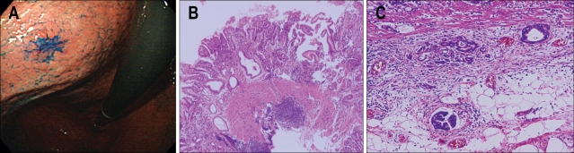
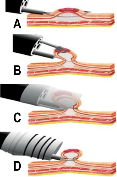
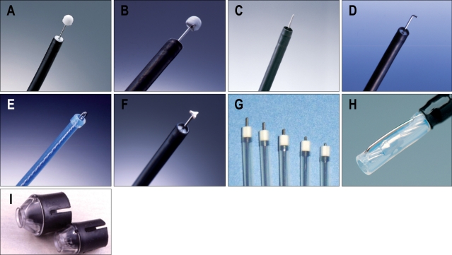
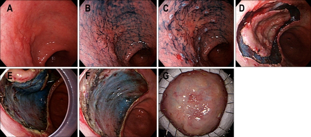
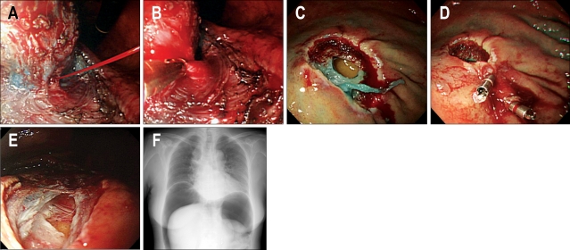
Early gastrointestinal cancers are defined as lesions limited to the mucosa or submucosa without invading the muscularis propria, regardless of the presence of lymph node metastases. Although the natural history of these diseases is basically alike worldwide, its management is quite different between the East and West; aggressive surgery is frequently adopted by Western surgeons, while less invasive techniques are adopted by Asian colleagues. These techniques include endoscopic mucosal resection and endoscopic submucosal dissection which are now accepted as treatments for early gastrointestinal cancers in selected cases. Recent advances in endoscopic detection and treatment techniques, especially in Japan and Korea, have prompted Western endoscopists to learn these techniques. This review addresses recent advances regarding endoscopic resections of early gastrointestinal cancers, which promoted its use in Western countries. In addition, prospective studies on endoscopic resection in Western countries are also described.
Keywords: Endoscopic resection; Gastrointestinal cancer; West.
Figures
References
-
- Rembacken BJ, Gotoda T, Fujii T, Axon AT. Endoscopic mucosal resection. Endoscopy. 2001;33:709–718. - PubMed
-
- Soetikno RM, Gotoda T, Nakanishi Y, Soehendra N. Endoscopic mucosal resection. Gastrointest Endosc. 2003;57:567–579. - PubMed
-
- Conio M, Ponchon T, Blanchi S, Filiberti R. Endoscopic mucosal resction. Am J Gastroenterol. 2006;101:653–663. - PubMed
-
- Fleischer D. Endoscopic mucosal resection: (not) made in the USA (so commonly). A dissection of the definition, technique, use, and controversies. Gastrointest Endosc. 2000;52:440–444. - PubMed
-
- Endoscopic Classification Review Group. Update on the Paris Classification of superficial neoplastic lesions in the digestive tract. Endoscopy. 2005;37:570–578. - PubMed
LinkOut - more resources
Full Text Sources
Other Literature Sources
