

In vivo imaging of alpha-synuclein in mouse cortex demonstrates stable expression and differential subcellular compartment mobility
- PMID: 20485674
- PMCID: PMC2868057
- DOI: 10.1371/journal.pone.0010589
In vivo imaging of alpha-synuclein in mouse cortex demonstrates stable expression and differential subcellular compartment mobility
Abstract
Background: Regulation of alpha-synuclein levels within cells is thought to play a critical role in Parkinson's Disease (PD) pathogenesis and in other related synucleinopathies. These processes have been studied primarily in reduced preparations, including cell culture. We now develop methods to measure alpha-synuclein levels in the living mammalian brain to study in vivo protein mobility, turnover and degradation with subcellular specificity.
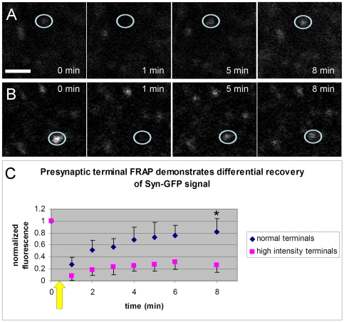
Methodology/principal findings: We have developed a system using enhanced Green Fluorescent Protein (GFP)-tagged human alpha-synuclein (Syn-GFP) transgenic mice and in vivo multiphoton imaging to measure alpha-synuclein levels with subcellular resolution. This new experimental paradigm allows individual Syn-GFP-expressing neurons and presynaptic terminals to be imaged in the living mouse brain over a period of months. We find that Syn-GFP is stably expressed by neurons and presynaptic terminals over this time frame and further find that different presynaptic terminals can express widely differing levels of Syn-GFP. Using the fluorescence recovery after photobleaching (FRAP) technique in vivo we provide evidence that at least two pools of Syn-GFP exist in terminals with lower levels of mobility than measured previously. These results demonstrate that multiphoton imaging in Syn-GFP mice is an excellent new strategy for exploring the biology of alpha-synuclein and related mechanisms of neurodegeneration.
Conclusions/significance: In vivo multiphoton imaging in Syn-GFP transgenic mice demonstrates stable alpha-synuclein expression and differential subcellular compartment mobility within cortical neurons. This opens new avenues for studying alpha-synuclein biology in the living brain and testing new therapeutics for PD and related disorders.
Conflict of interest statement
Figures


Similar articles
-
Presynaptic alpha-synuclein aggregation in a mouse model of Parkinson's disease.J Neurosci. 2014 Feb 5;34(6):2037-50. doi: 10.1523/JNEUROSCI.2581-13.2014. J Neurosci. 2014. PMID: 24501346 Free PMC article.
-
Longitudinal live imaging of retinal α-synuclein::GFP deposits in a transgenic mouse model of Parkinson's Disease/Dementia with Lewy Bodies.Sci Rep. 2016 Jul 8;6:29523. doi: 10.1038/srep29523. Sci Rep. 2016. PMID: 27389831 Free PMC article.
-
A novel α-synuclein-GFP mouse model displays progressive motor impairment, olfactory dysfunction and accumulation of α-synuclein-GFP.Neurobiol Dis. 2013 Aug;56:145-55. doi: 10.1016/j.nbd.2013.04.017. Epub 2013 Apr 30. Neurobiol Dis. 2013. PMID: 23643841
-
Neurotoxic conversion of beta-synuclein: a novel approach to generate a transgenic mouse model of synucleinopathies?J Neurol. 2009 Aug;256 Suppl 3:286-92. doi: 10.1007/s00415-009-5246-8. J Neurol. 2009. PMID: 19711118 Review.
-
Implication of Alpha-Synuclein Phosphorylation at S129 in Synucleinopathies: What Have We Learned in the Last Decade?J Parkinsons Dis. 2016;6(1):39-51. doi: 10.3233/JPD-160779. J Parkinsons Dis. 2016. PMID: 27003784 Free PMC article. Review.
Cited by
-
The physiological role of α-synuclein and its relationship to Parkinson's Disease.J Neurochem. 2019 Sep;150(5):475-486. doi: 10.1111/jnc.14810. Epub 2019 Jul 28. J Neurochem. 2019. PMID: 31269263 Free PMC article. Review.
-
Development and screening of contrast agents for in vivo imaging of Parkinson's disease.Mol Imaging Biol. 2013 Oct;15(5):585-95. doi: 10.1007/s11307-013-0634-y. Mol Imaging Biol. 2013. PMID: 23624948
-
Protein degradation pathways in Parkinson's disease: curse or blessing.Acta Neuropathol. 2012 Aug;124(2):153-72. doi: 10.1007/s00401-012-1004-6. Epub 2012 Jun 29. Acta Neuropathol. 2012. PMID: 22744791 Free PMC article. Review.
-
α-Synuclein inhibits intersynaptic vesicle mobility and maintains recycling-pool homeostasis.J Neurosci. 2012 Jul 25;32(30):10129-35. doi: 10.1523/JNEUROSCI.0535-12.2012. J Neurosci. 2012. PMID: 22836248 Free PMC article.
-
Direct detection of alpha synuclein oligomers in vivo.Acta Neuropathol Commun. 2013 May 9;1:6. doi: 10.1186/2051-5960-1-6. Acta Neuropathol Commun. 2013. PMID: 24252244 Free PMC article.
References
-
- Spillantini MG, Schmidt ML, Lee VM, Trojanowski JQ, Jakes R, et al. Alpha-synuclein in lewy bodies. Nature. 1997;388(6645):839–40. - PubMed
-
- Farrer M, Kachergus J, Forno L, Lincoln S, Wang DS, et al. Comparison of kindreds with parkinsonism and alpha-synuclein genomic multiplications. Ann Neurol. 2004;55(2):174–9. - PubMed
-
- Gomez-Santos C, Barrachina M, Gimenez-Xavier P, Dalfo E, Ferrer I, et al. Induction of C/EBP beta and GADD153 expression by dopamine in human neuroblastoma cells: relationship with alpha-synuclein increase and cell damage. Brain Res Bull. 2005;65(1):87–95. - PubMed
-
- Yang YX, Latchman DS. Nurr1 transcriptionally regulates the expression of alpha-synuclein. Neuroreport. 2008;19(8):867–71. - PubMed
Publication types
MeSH terms
Substances
Grants and funding
- T32AG000222/AG/NIA NIH HHS/United States
- T32 AG000222/AG/NIA NIH HHS/United States
- T32 NS048005/NS/NINDS NIH HHS/United States
- R01 NS063963/NS/NINDS NIH HHS/United States
- NS038372/NS/NINDS NIH HHS/United States
- R01 AG018440/AG/NIA NIH HHS/United States
- NS063963/NS/NINDS NIH HHS/United States
- R37 AG018440/AG/NIA NIH HHS/United States
- T32NS048005/NS/NINDS NIH HHS/United States
- P50 NS038372/NS/NINDS NIH HHS/United States
- AG022074/AG/NIA NIH HHS/United States
- AG18440/AG/NIA NIH HHS/United States
- P01 AG022074/AG/NIA NIH HHS/United States
LinkOut - more resources
Full Text Sources
Molecular Biology Databases
Miscellaneous
