. 2010 Jun 9;10(6):1971-8.
doi: 10.1021/nl101262u.

Structural DNA nanotechnology: growing along with Nano Letters

Affiliations

Structural DNA nanotechnology: growing along with Nano Letters

Nadrian C Seeman. Nano Lett. .

Abstract

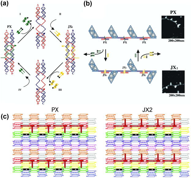
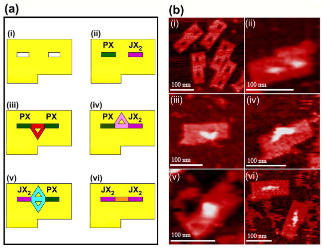
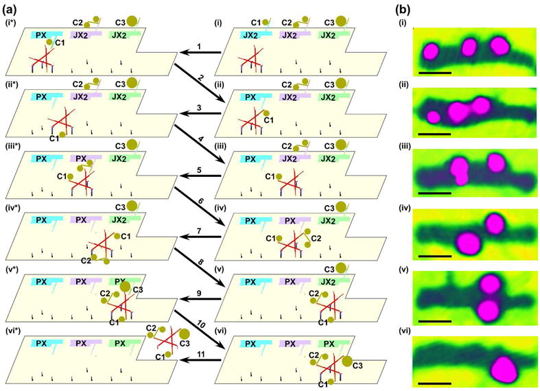
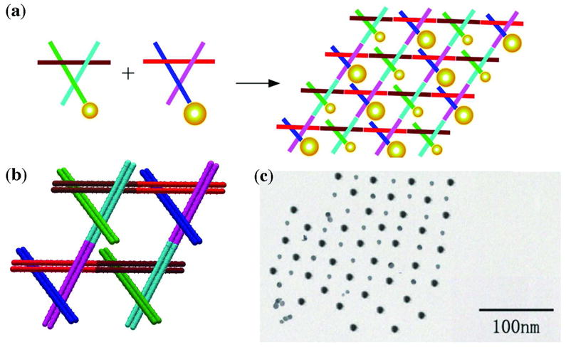
During the past decade, the field of structural DNA nanotechnology has grown enormously, not only in the number of its participants but also qualitatively in its capabilities. A number of goals evident in 2001 have been achieved: These include the extension of self-assembled crystalline systems from 2D to 3D and the achievement of 2D algorithmic assembly. A variety of nanoscale walking devices have been developed. A key unanticipated development was the advent of DNA origami, which has vastly expanded the scale of addressable DNA structures. Nanomechanical devices have been incorporated into 2D arrays, and into 2D origami structures, as well, leading to capture systems and to a nanomechanical assembly line. DNA has been used to scaffold non-DNA species, so that one of its key goals has been achieved. Biological replication of DNA nanostructures with simple topologies has also been accomplished. The increase in the number of participants in the enterprise holds great promise for the coming decade.


Figures

Figure 1
Figure 1. A Stereoscopic Image of the Tensegrity Triangle
The three unique strands are shown in individual colors. The blue strands are on the periphery of the triangle, the yellow strands form continuous helices and the red strand is a cyclic covalent strand containing a single nick distal to any crossover points.
Figure 2
Figure 2. The Lattice Formed by the Tensegrity Triangles
(a) The Surroundings of an Individual Triangle. This stereoscopic simplified image distinguishes the three independent directions by the colors (red, green and yellow) of their base pairs. Thus, the central triangle is shown flanked by three other pairs of triangles in the three differently colored directions. (b) The Rhombohedral Cavity Formed by the Tensegrity Triangles. This stereoscopic projection shows seven of the eight tensegrity triangles that comprise the corners of the rhombohedron. The outline of the cavity is shown in white. The red triangle at the back connects through one edge each to the three yellow triangles that lie in a plane somewhat closer to the viewer. The yellow triangles are connected through two edges each to two different green triangles that are in a plane even nearer the viewer. A final triangle that would cap the structure has been omitted for clarity. This triangle would be directly above the red triangle, and would be even closer to the viewer than the green triangles.
Figure 3
Figure 3. Atomic Force Micrographs of DNA Origami Constructs
(a) A Smiley Face. The scaffold strand, bound to helper strands, zigzags back and forth from left to right, yielding the structure seen. (b) A Map of the Western Hemisphere. This image demonstrates dramatically the addressability of the DNA array. Each of the white pixels is made by a small DNA double helical domain roughly normal to the surface.
Figure 4
Figure 4. Seeded and Chaperoned Growth of Sierpinski Triangles
The upper panel shows an AFM image of a Sierpinski triangle that is seeded by a DNA origami on the left. The lower panel is an interpretation. The tiles that flank the rectangle on the top and the bottom provide extra control on the growth of the image. The scale bar is 100 nm, and the arrow indicates where analysis stopped following change in width of the sample.
Figure 5
Figure 5. DNA-Based Nanomechanical Devices
(a) The Machine Cycle of a PX-JX2 Device. Starting with the PX state on the left, the green set strands are removed by their complements (Process I) to leave an unstructured frame. The addition of the yellow set strands (process II) converts the frame to the JX2 state, in which the top and bottom domains are rotated a half turn relative to their arrangement in the PX conformation. Processes III and IV reverse this process to return to the PX structure. (b) AFM Demonstration of the Operation of the Device. A series of DNA trapezoids are connected by devices. In the PX state, the trapezoids are in a parallel arrangement, but when the system is converted to the JX2 state, they are in a zigzag arrangement. (c) Insertion of a Device Cassette into a 2D Array. The eight TX tiles that form the array are shown in differently colored outlined tiles. For clarity the cohesive ends are shown to be the same geometrical shape, although they all contain different sequences. The domain connecting the cassette to the lattice is not shown. The cassette and reporter helix are shown as red filled components; the black marker tile is labeled ‘M’ and is shown with a black filled rectangle representing the domain of the tile that protrudes from the rest of the array. Both the cassette and the marker tile are rotated about 103° from the other components of the array (three nucleotides rotation). The PX arrangement is shown at the left and the JX2 arrangement is on the right. Note that the reporter hairpin points towards the marker tile in the PX state, but points away from it in the JX2 state.
Figure 6
Figure 6. Schematics (a) and Atomic Force Micrographs (b) of the Origami Arrays and Capture Molecules
Panel i of (a) illustrates the origami array containing slots for the cassettes and a notch to enable recognition of orientation; the slots and notches are visible in the AFM in (b). Panels ii show the cassettes in place; the color coding in (a) used throughout the schematics is green for the PX state and violet for the JX2 state; the presence of the cassettes is evident in the AFM image in (b). Panels iii illustrate the PX-PX state which captures a triangle pointing towards the notch in the schematic (a) and in the AFM image (b). Panels iv illustrate the PX-JX2 state (a), containing a triangle that points away from the notch, which is evident in the AFM image (b). Panels v illustrate the JX2- PX state which captures a diamond-shaped molecule (a); its shape is visible in the AFM image (b). Panels vi show the linear molecule captured by the JX2-JX2 state, both schematically (a) and in the AFM image (b).
Figure 7
Figure 7. The Steps in the Assembly Line Construction of a Triple Addition Product
Schematics are shown in (a) and atomic force micrographs of the right column of (a) are shown in (b). AFM was performed by tapping in air; this mode of AFM results in only the nanoparticles and the origami being visible, and the individual nanoparticle components are not resolved from each other. Owing to the washing procedures between steps, the AFM images are not of the same individual assembly line. Panel i illustrates the origami array with cassettes and walker in the starting position. The cassettes are set to the default JX2 state, with the arms pointing away from the walker pathway. Different cargoes on the arms (5 nm Au on cassette 1, a linked 5 nm Au pair on cassette 2, and a 10 nm Au on cassette 3) are visible both schematically (a) and in the AFM (b). Step 1 shows cassette 1 switched from the JX2 state to the PX state, bringing cargo 1 close to the walker. Step 2 illustrates the addition of cargo 1 from cassette 1 to the walker by DNA branch migration; the movement of cargo 1 is evident in the AFM (ii). Step 3 shows the walker with cargo 1 walking the 1st half-step along the pathway; step 4 illustrates the walker with cargo 1 walking the 2nd half-step, positioning itself near cassette 2, which is visible both schematically and in the AFM (iii). Step 5 shows cassette 2 is switched from the JX2 state to the PX state, bringing cargo 2 close to the walker. Step 6 illustrates the addition of cargo 2 from cassette 2 to the walker by branch migration; the addition of cargo 2 is evident in the AFM (iv). Step 7 shows the walker with cargo 1 and cargo 2 walking the 3rd half-step along the pathway. Step 8 illustrates the walker with both cargo 1 and cargo 2 walking the 4th half-step to be close to cassette 3; the walking is clearly visible in the AFM (v). Step 9 shows cassette 3 switched from the JX2 state to the PX state, bringing cargo 3 close to the walker. Step 10 illustrates the addition of cargo 3 from cassette 3 to the walker by branch migration; the addition of cargo 3 is visible in the AFM (vi). Step 11 shows the walker with all three cargo components released from the origami. All scale bars are 50 nm.
Figure 8
Figure 8. Organizing Gold Nanoparticles with a 2D Motif
(a) Two different 3D-DX motifs, containing 5 nm or 10 nm particles on one end of a propagation direction yield a checkerboard nanoparticle array. (b) The two 3D-DX motifs in greater detail assembled to form a 2D array. (c) A TEM image showing a checkerboard array of gold nanoparticles.

References

    1. Seeman NC. J Theor Biol. 1982;99:237–247. - PubMed
    1. Seeman NC. NanoLetters. 2001;1:22–26.
    1. Chen J, Seeman NC. Nature. 1991;350:631–633. - PubMed
    1. Zhang Y, Seeman NC. J Am Chem Soc. 1994;116:1661–1669.
    1. Winfree E, Liu F, Wenzler LA, Seeman NC. Nature. 1998;394:539–544. - PubMed

Publication types