. 2010 Oct;16(10):3023-31.
doi: 10.1089/ten.TEA.2010.0181.

Regeneration of dental-pulp-like tissue by chemotaxis-induced cell homing

Affiliations

Regeneration of dental-pulp-like tissue by chemotaxis-induced cell homing

Jin Y Kim et al. Tissue Eng Part A. 2010 Oct.

Abstract

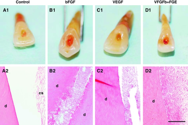
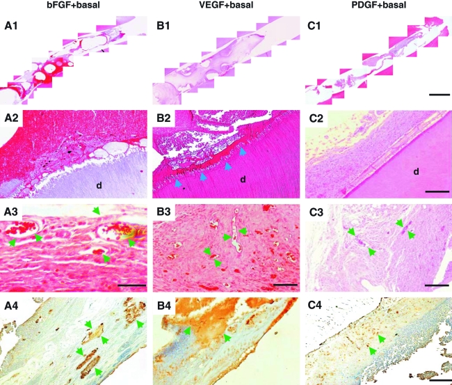
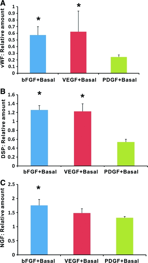
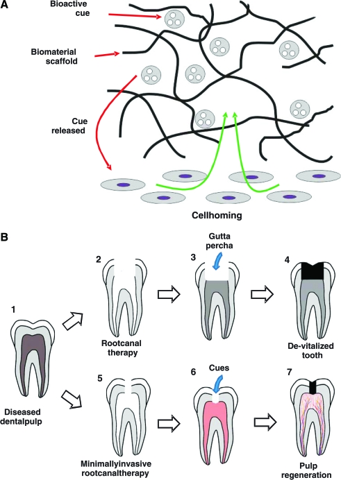
Tooth infections or injuries involving dental pulp are treated routinely by root canal therapy. Endodontically treated teeth are devitalized, susceptible to re-infections, fractures, and subsequent tooth loss. Here, we report regeneration of dental-pulp-like tissue by cell homing and without cell transplantation. Upon in vivo implantation of endodontically treated real-size, native human teeth in mouse dorsum for the tested 3 weeks, delivery of basic fibroblast growth factor and/or vascular endothelial growth factor (bFGF and/or VEGF) yielded re-cellularized and revascularized connective tissue that integrated to native dentinal wall in root canals. Further, combined delivery of bFGF, VEGF, or platelet-derived growth factor (PDGF) with a basal set of nerve growth factor (NGF) and bone morphogenetic protein-7 (BMP7) generated cellularized and vascularized tissues positive of VEGF antibody staining and apparent neo-dentin formation over the surface of native dentinal wall in some, but not all, endodontically treated teeth. Newly formed dental pulp tissue appeared dense with disconnected cells surrounded by extracellular matrix. Erythrocyte-filled blood vessels were present with endothelial-like cell lining. Reconstructed, multiple microscopic images showed complete fill of dental-pulp-like tissue in the entire root canal from root apex to pulp chamber with tissue integration to dentinal wall upon delivery of bFGF, VEGF, or PDGF with a basal set of NGF and BMP7. Quantitative ELISA showed that combinatory delivery of bFGF, VEGF, or PDGF with basal NGF and BMP7 elaborated von Willerbrand factor, dentin sialoprotein, and NGF. These findings represent the first demonstration of regenerated dental-pulp-like tissue in endodontically treated root canals of real-size, native human teeth. The present chemotaxis-based approach has potent cell homing effects for re-cellularization and revascularization in endodontically treated root canals in vivo, although in an ectopic model. Regeneration of dental pulp by cell homing, rather than cell delivery, may accelerate clinical translation.


Figures

FIG. 1.
FIG. 1.
Re-cellularization and revascularization of endodontically treated root canals in real-size human teeth. Clinically extracted human teeth were treated endodontically as in patients, with the exception of filling with gutta percha, a bioinert thermoplastic material. Instead, collage scaffolds were implanted in endodontically treated root canals with or without delivery of basic fibroblast growth factor (bFGF) and/or vascular endothelial growth factor (VEGF) followed by 3-week in vivo implantation. (A1) Endodontically treated root canals with collagen scaffold alone showed pale access opening. (A2) Microscopically, root canals with collagen scaffold alone showed residual collagen scaffold (cs) that is adjacent to native dentin (d), but little cell ingrowth. (B1) bFGF delivery in collagen scaffold yielded red pigmentation. (B2) Corresponding micrograph showing re-cellularization of endodontically treated root canal with abundant cells and some extracellular matrix that integrated with the wall of native dentin (d). (C1) VEGF delivery yielded re-cellularization with connective tissue within root canal (C2). Separation of soft tissue with dentinal wall is likely due to artifacts in histology processing, in consideration of tissue integration with dentin in other groups and also in Figure 2 below. Combined bFGF and VEGF delivery also generated red pigmentation (D1) and abundant cells within root canal (D2). Scale bars (A2, B2, C2, D2): 500 μm. Color images available online at www.liebertonline.com/ten.
FIG. 2.
FIG. 2.
Combinatory cytokine delivery and chemotactic effects on pulp regeneration from host endogenous cells. (A1) Combined delivery of bFGF with basal cytokines of nerve growth factor (NGF) and bone morphogenetic protein-7 (BMP7) yielded re-cellularization in the entire endodontically treated root canal as shown by reconstructed, multiple microscopic images from root apex to pulp chamber. (A2) Close-up image showing abundant cells and integration with native dentin (d) in endodontically treated root canal. (A3) Higher magnification showing erythrocyte-filled blood vessels (arrows) in connective tissue. (A4) Scattered islands of positive VEGF antibody staining (arrows). (B1) Combined delivery of VEGF with basal cytokines of NGF and BMP7 induced re-cellularization in the entire endodontically treated root canal. (B2) Connective tissue with abundant cells integrated with native dentin (d), including a layer of dentin-like tissue between abundant cells and dentinal wall (arrows). (B3) Multiple blood-vessel-like structures in regenerated connective tissue (arrows). (B4) Positive VEGF antibody staining. Arrows indicate positive VEGF staining. (C1) Combined delivery of platelet-derived growth factor (PDGF) with basal cytokines of NGF and BMP7 yielded fully regenerated tissues in the entire endodontically treated root canal. (C2) Abundant cells in connective tissue that integrated to native dentin (d). (C3) Blood-vessel-like structures (arrows) in connective tissue. (C4) VEGF antibody staining. Arrows indicate positive VEGF staining. Scale bars (A1, B1, C1): 1 mm; (A2, B2, C2): 100 μm; (A3): 100 μm; (B3, C3): 300 μm; (A4, B4, C4): 300 μm. Color images available online at www.liebertonline.com/ten.
FIG. 3.
FIG. 3.
ELISA of von Willerbrand factor (vWF), dentin sialoprotein (DSP), and NGF in regenerated dental-pulp-like tissue relative to collagen scaffold only group. (A) Codelivery of bFGF or VEGF with basal cytokines of NGF and BMP7 yielded significantly more vWF than PDGF with NGF and BMP7. (B) Codelivery of bFGF or VEGF with basal cytokines of NGF and BMP7 yielded significantly more DSP than PDGF with NGF and BMP7. (C) Codelivery of bFGF or VEGF with basal cytokines of NGF and BMP7 yielded significantly more NGF than PDGF with NGF and BMP7. n = 6; *p < 0.05. Color images available online at www.liebertonline.com/ten.
FIG. 4.
FIG. 4.
Schemes of cell homing for dental pulp regeneration and clinical translation. (A) Bioactive cues can be adsorbed, tethered, or encapsulated in biomaterial scaffolds. Upon release of bioactive cues, such as from endodontically treated root canals in this work, local and/or systemic cells, including stem/progenitor cells, can be homed in vivo into an anatomic compartment, which in this case, is root canal that serves as a native scaffold. Current root canal treatment of diseased dental pulp (B1) necessitates removal of substantial enamel and dentin structures because obturation of gutta percha requires unobstructed access (B2, B3), yet leading to a de-vitalized tooth (B4). We propose that a diseased dental pulp (B1) can be treated with a revised, minimally invasive root canal therapy (B5) on the basis that delivery of injectable bioactive cues does not require unobstructed access to pulp chamber and root canal (B6). Although residual inflammation in endodontically treated root canal and peri-apical region are anticipated to present challenges for pulp regeneration, chemotaxis-induced angiogenesis as shown in the present may provide the potential for native defense mechanisms that may counteract residue infection in the root canal (B6), leading to a vital tooth with regenerated dental pulp (B7). Color images available online at www.liebertonline.com/ten.

Similar articles

Cited by

References

    1. Ten Cate A.R. Dentin and pulp. In: Ten Cate A.R., editor. Oral Histology. London: Mosby; 1998. pp. 246–278.
    1. Ingle J.I. Bakland L.K. Structure and function of the dentin-pulp complex. In: Ingle J.I., editor; Bakland L.K., editor. Endodontics. Hamilton, Ontario, London: BC Decker, Inc.; 2002. pp. 121–143.
    1. Dammaschke T. Steven D. Kaup M. Ott K.H. Long-term survival of root-canal-treated teeth: a retrospective study over 10 years. J Endod. 2003;29:638. - PubMed
    1. Andreasen J.O. Farik B. Munksgaard E.C. Long-term calcium hydroxide as a root canal dressing may increase risk of root fracture. Dent Traumatol. 2002;18:134. - PubMed
    1. Caplan D.J. Cai J. Yin G. White B.A. Root canal filled versus non-root canal filled teeth: a retrospective comparison of survival times. J Public Health Dent. 2005;65:90. - PubMed

Publication types

MeSH terms

LinkOut - more resources