. 2010 Feb;75(4):924-41.
doi: 10.1111/j.1365-2958.2009.07027.x.

Control of effector export by the Pseudomonas aeruginosa type III secretion proteins PcrG and PcrV

Affiliations

Control of effector export by the Pseudomonas aeruginosa type III secretion proteins PcrG and PcrV

Pei-Chung Lee et al. Mol Microbiol. 2010 Feb.

Erratum in

  • Mol Microbiol. 2010 Mar;75(5):1325

Abstract

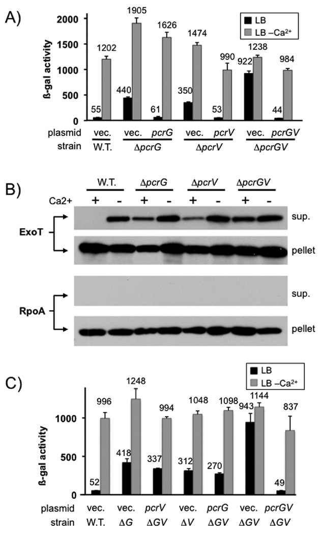
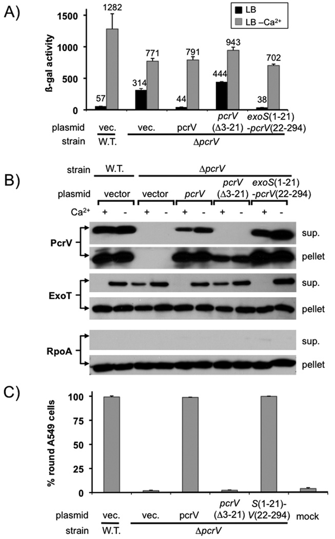
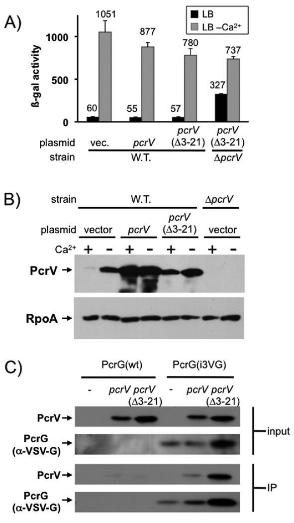
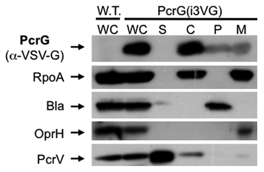
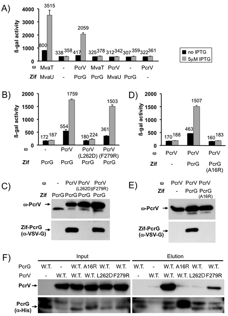
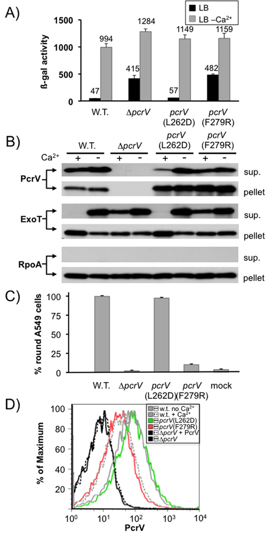
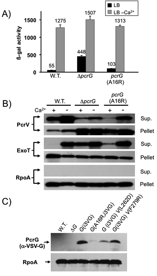
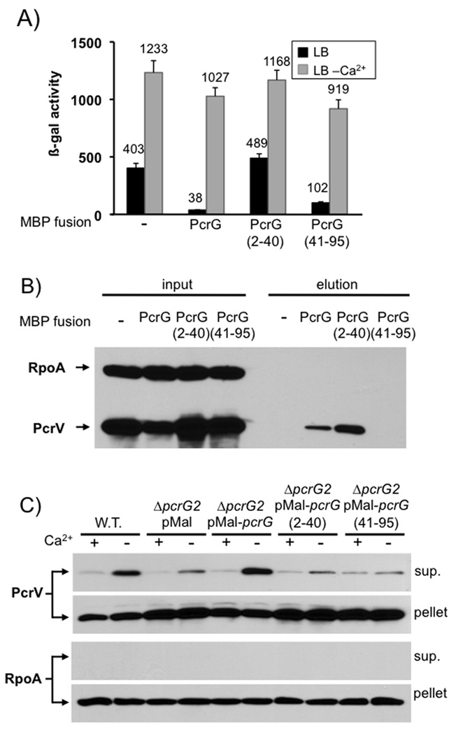
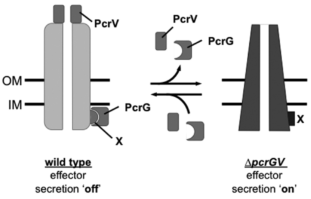
Pseudomonas aeruginosa uses a type III secretion system to inject protein effectors into a targeted host cell. Effector secretion is triggered by host cell contact. How effector secretion is prevented prior to cell contact is not well understood. In all secretion systems studied to date, the needle tip protein is required for controlling effector secretion, but the mechanism by which needle tip proteins control effector secretion is unclear. Here we present data that the P. aeruginosa needle tip protein, PcrV, controls effector secretion by assembling into a functional needle tip complex. PcrV likely does not simply obstruct the secretion channel because the pore-forming translocator proteins can still be secreted while effector secretion is repressed. This finding suggests that PcrV controls effector secretion by affecting the conformation of the apparatus, shifting it from the default, effector secretion 'on' conformation, to the effector secretion 'off' conformation. We also present evidence that PcrG, which can bind to PcrV and is also involved in controlling effector export, is cytoplasmic and that the interaction between PcrG and PcrV is not required for effector secretion control by either protein. Taken together, these data allow us to propose a working model for control of effector secretion by PcrG and PcrV.


Figures

Fig. 1
Fig. 1. pcrG and pcrV mutant phenotype
(A) The effect of deleting pcrG and/or pcrV was assayed by measuring β-galactosidase activity of P. aeruginosa PAO1 in which the chromosomal copy of exoS has been replaced by a tandem set of translationally coupled reporters (GFP-lacZ). Where indicated, a wild-type copy of pcrG or pcrV was supplied in trans from a plasmid (vec = vector control). (B) export of ExoT was measured by RECC assay. Supernatant and pellet fractions are indicated. The RNA polymerase alpha subunit (RpoA) served as fractionation control. (C) a ΔpcrGV double null mutant was complemented by either expressing pcrG alone, pcrV alone or both open reading frames together. The pcrG plasmid was induced with 10µM IPTG, the pcrV plasmid with 125 µM IPTG and the pcrGV plasmid with 10µM IPTG. The success of the complementation was monitored using the chromosomal exoS reporter fusion described in (A).
Fig. 2
Fig. 2. PcrV requires an intact secretion signal to control effector export
(A) a pcrV deletion mutant was complemented with either wild-type pcrV, a pcrV deletion mutant lacking codons 3–21, or a fusion in which codons 1–21 of exoS were fused to codons 22–294 of pcrV. The ability of the indicated ORF to complement the pcrV null mutant phenotype was monitored using a lacZ reporter inserted in the exoS locus (A) or by RECC assay (B). The ability of the indicated open reading frames to complement the defect in cytotoxicity of a pcrV null mutant was assayed by monitoring rounding of A549 cells after 5 hours of infection (MOI 10)(C).
Fig. 3
Fig. 3. Overexpression of PcrV or PcrV (Δ3–21)
PcrV or the signal-sequenceless version PcrV(Δ3–21) were overexpressed in a wild-type strain background either in the presence or absence of calcium. The effect on the export of effectors was monitored using a lacZ reporter inserted in the exoS locus (A). Overexpression of PcrV or PcrV (Δ3–21) was confirmed by western blot on cell pellets of overexpressing strains (B, RpoA served as loading control). (C) Wild-type PcrV or a mutant lacking the export signal (Δ3–21) were expressed from a plasmid in a strain in which the chromosomal copy of pcrV had been deleted and in which expression of the type III secretion genes was uniformly up-regulated (ΔexsE). Where indicated, the strain also harbored a chromosomal copy of pcrG, which had been modified to express an internally VSV-G tagged version of the protein, PcrG(i3VG). Both input cell-lysates and output fractions after immunoprecipitation with the anti-VSV-G tag antibody were separated by SDS-PAGE and probed for the presence of PcrV or the VSV-G-tagged PcrG as indicated.
Fig. 4
Fig. 4. Co-immunoprecipitation of PcrG and PcrV
A functional, VSV-G-tagged version of PcrG (PcrG(i3VG)) was used to determine the localization of PcrG in the cell. Bacteria were fractionated using the lysozyme/EDTA method. Fractions are indicated above each lane (wc = whole cell, S = supernatant, C = cytoplasm, P = periplasm, M = membrane). Fractionation was controlled using markers for secreted proteins (PcrV), membrane proteins (OprH), periplasmic proteins (beta-lactamase) and the cytoplasm (RpoA).
Fig. 5
Fig. 5. The interaction between PcrG and PcrV
The interaction between PcrG and PcrV was assayed by two-hybrid analysis. PcrV was fused to the omega-subunit of RNA polymerase (ω). PcrG was fused to the monomeric DNA binding protein Zif. Interaction of the fusion proteins results in recruitment of RNA polymerase to a test promoter, which can be readily assayed by β-galactosidase assay. PcrG and PcrV interact with each other and not the unrelated histone-like proteins MvaT and MvaU (A). The effect of mutations in PcrV (B) or PcrG (D) on the interaction was monitored by using the same two-hybrid system, the only modification being that the Zif-fusion had been modified by inserting a VSV-G tag into the linker between Zif and PcrG. Production of the ω and Zif fusion proteins was monitored by western blot (C, E). Interaction data was confirmed by pull-down in E. coli (F). PcrV and a His-tagged version of PcrG, as well as mutants of either protein, were co-expressed in E. coli BL21. Co-purification of PcrG and PcrV was monitored by detecting PcrV in the elution fraction after purification.
Fig. 6
Fig. 6. Phenotypic characterization of pcrV point mutants
The effect of point mutations L262D and F279R in pcrV was assayed by β-galactosidase assay using a chromosomal lacZ reporter inserted in the exoS locus (A). It was also examined by RECC assay (B). Secretion of PcrV, ExoT and RpoA (control) was monitored by Western blot (the respective protein detected is indicated to the left of each blot, the fraction (cell pellet or supernatant) is indicated to the right). The ability to intoxicate cells was determined by monitoring rounding of A549 cells after 5 hours of infection (MOI10). Surface localization of PcrV was monitored by FACS using an affinity-purified anti-PcrV antiserum and an anti-rabbit IgG APC-conjugated secondary antibody. To normalize expression of PcrV, all strains were grown in the absence of calcium, except the wild-type, which was grown both in the absence and presence of calcium (indicated in the legend). Purified PcrV protein was added to one sample of the pcrV null mutant before performing the antibody staining procedure in order to rule out the possibility that secreted protein being non-specifically cross-linked to the cells interfered with our assay.
Fig. 7
Fig. 7. Phenotypic characterization of pcrG point mutants
The effect of mutating the chromosomal copy of pcrG was assayed by β-galactosidase assay, using a lacZ reporter inserted in the exoS locus (A), or by RECC assay (B). Proteins detected in the RECC assay are indicated to the left of each blot, the fraction to the right. (C) Stability of PcrG was assayed by modifying the chromosomal copy of pcrG to express an internally VSV-G tagged version of PcrG (PcrG(i3VG)). The western blot was performed on cell pellets of bacteria grown under T3SS inducing (low calcium) conditions.
Fig. 8
Fig. 8. PcrV-binding and regulatory functions of PcrG can be separated
Full length PcrG or truncated versions (aa 2–40 or 41–95) were fused to the C-terminus of MBP and expressed in a mutant of P. aeruginosa lacking pcrG. The ability to control effector export was monitored by β-galactosidase assay using a lacZ reporter inserted into the chromosomal exoS locus (A). The ability of the MBP-fusions to bind to PcrV was determined by pull-down from cytoplasmic extracts using an amylose-resin, followed by Western blot analysis (B). Secretion of PcrV was analyzed by RECC assay (C). The genotype and plasmid is noted above each set of lanes. Supernatant and cell pellet fractions, as well as the protein being detected are listed to the side of the relevant panel.
Fig. 9
Fig. 9. Model of PcrV function in type III secretion regulation
We propose that the T3SS is in the effector secretion ‘on’ position in the absence of PcrG and PcrV (e.g. in the case of the pcrGV null mutant). The assembly of PcrV at the needle-tip (and perhaps binding of PcrG to a cytoplasmic component(s) of the T3SS [X]) shifts the conformation of the apparatus to the effector secretion ‘off’ conformation.

Similar articles

Cited by

References

    1. Bergman T, Hakansson S, Forsberg A, Norlander L, Macellaro A, Backman A, Bolin I, Wolf-Watz H. Analysis of the V antigen lcrGVH-yopBD operon of Yersinia pseudotuberculosis: evidence for a regulatory role of LcrH and LcrV. J Bacteriol. 1991;173:1607–1616. - PMC - PubMed
    1. Bleves S, Soscia C, Nogueira-Orlandi P, Lazdunski A, Filloux A. Quorum sensing negatively controls type III secretion regulon expression in Pseudomonas aeruginosa PAO1. J Bacteriol. 2005;187:3898–3902. - PMC - PubMed
    1. Blocker AJ, Deane JE, Veenendaal AK, Roversi P, Hodgkinson JL, Johnson S, Lea SM. What's the point of the type III secretion system needle? Proc Natl Acad Sci U S A. 2008;105:6507–6513. - PMC - PubMed
    1. Botteaux A, Sory MP, Biskri L, Parsot C, Allaoui A. MxiC is secreted by and controls the substrate specificity of the Shigella flexneri type III secretion apparatus. Mol Microbiol. 2008 - PubMed
    1. Broms JE, Forslund AL, Forsberg A, Francis MS. PcrH of Pseudomonas aeruginosa is essential for secretion and assembly of the type III translocon. J Infect Dis. 2003;188:1909–1921. - PubMed

Publication types

MeSH terms

Substances