

Antibodies against gonadotropin-releasing hormone (GnRH) and destruction of enteric neurons in 3 patients suffering from gastrointestinal dysfunction
- PMID: 20487533
- PMCID: PMC2885307
- DOI: 10.1186/1471-230X-10-48
Antibodies against gonadotropin-releasing hormone (GnRH) and destruction of enteric neurons in 3 patients suffering from gastrointestinal dysfunction
Abstract
Background: Antibodies against gonadotropin-releasing hormone (GnRH) and gastrointestinal dysmotility have been found after treatment with GnRH analogues. The aim of this study was to examine the presence of such antibodies in patients with dysmotility not subjected to GnRH treatment and study the anti-GnRH antibody effect on enteric neurons viability in vitro.
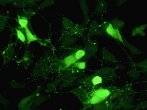
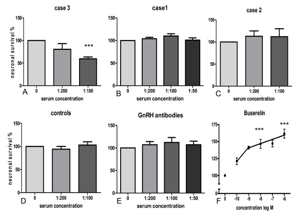
Methods: Plasma and sera from 3 patients suffering from either enteric dysmotility, irritable bowel syndrome (IBS) or gastroparesis were analysed for C-reactive protein (CRP), and for GnRH antibodies and soluble CD40 by ELISA methods. Primary cultures of small intestinal myenteric neurons were prepared from rats. Neuronal survival was determined after the addition of sera either from the patients with dysmotility, from healthy blood donors, antiserum raised against GnRH or the GnRH analogue buserelin. Only for case 1 a full-thickness bowel wall biopsy was available for immunohistochemical analysis.
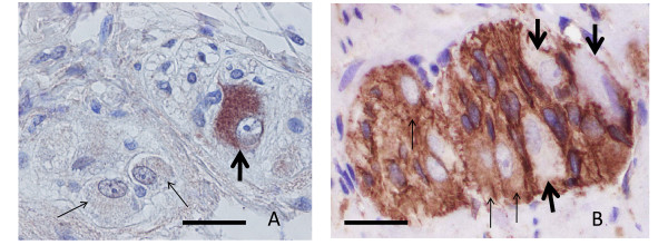
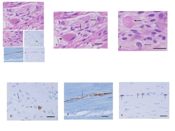
Results: All 3 patients expressed antibodies against GnRH. The antibody titer correlated to the levels of CD40 (rs = 1.000, p < 0.01), but not to CRP. Serum from case 3 with highest anti-GnRH antibody titer, and serum concentrations of sCD40 and CRP, when added to cultured rat myenteric neurons caused remarkable cell death. In contrast, serum from cases 1 and 2 having lower anti-GnRH antibody titer and lower sCD40 levels had no significant effect. Importantly, commercial antibodies against GnRH showed no effect on neuron viability whereas buserelin exerted a protective effect. The full-thickness biopsy from the bowel wall of case 1 showed ganglioneuritis and decrease of GnRH and GnRH receptor.
Conclusion: Autoantibodies against GnRH can be detected independently on treatment of GnRH analogue. Whether the generation of the antibody is directly linked to neuron degeneration and chronic gastrointestinal symptoms in patients with intestinal dysmotility, remains to be answered.
Figures

Similar articles
-
Depletion of enteric gonadotropin-releasing hormone is found in a few patients suffering from severe gastrointestinal dysmotility.Scand J Gastroenterol. 2012 Oct;47(10):1165-73. doi: 10.3109/00365521.2012.706826. Epub 2012 Jul 27. Scand J Gastroenterol. 2012. PMID: 22835010
-
Gonadotropin-releasing hormone analog buserelin causes neuronal loss in rat gastrointestinal tract.Cell Tissue Res. 2013 Mar;351(3):521-34. doi: 10.1007/s00441-012-1534-1. Epub 2012 Dec 20. Cell Tissue Res. 2013. PMID: 23254679
-
Patients with irritable bowel syndrome and dysmotility express antibodies against gonadotropin-releasing hormone in serum.Neurogastroenterol Motil. 2011 Nov;23(11):1000-6, e459. doi: 10.1111/j.1365-2982.2011.01744.x. Epub 2011 Jun 30. Neurogastroenterol Motil. 2011. PMID: 21714833
-
Gonadotropin-Releasing Hormone and Its Physiological and Pathophysiological Roles in Relation to the Structure and Function of the Gastrointestinal Tract.Eur Surg Res. 2016;57(1-2):22-33. doi: 10.1159/000445717. Epub 2016 Apr 19. Eur Surg Res. 2016. PMID: 27089503 Review.
-
Gonadotropin-Releasing Hormone and Its Role in the Enteric Nervous System.Front Endocrinol (Lausanne). 2017 Jun 7;8:110. doi: 10.3389/fendo.2017.00110. eCollection 2017. Front Endocrinol (Lausanne). 2017. PMID: 28638366 Free PMC article. Review.
Cited by
-
Sirtuin-3 Is Expressed by Enteric Neurons but It Does not Play a Major Role in Their Regulation of Oxidative Stress.Front Cell Neurosci. 2016 Mar 22;10:73. doi: 10.3389/fncel.2016.00073. eCollection 2016. Front Cell Neurosci. 2016. PMID: 27047337 Free PMC article.
-
Antibodies against gonadotropin-releasing hormone (GnRH) in patients with diabetes mellitus is associated with lower body weight and autonomic neuropathy.BMC Res Notes. 2013 Aug 17;6:329. doi: 10.1186/1756-0500-6-329. BMC Res Notes. 2013. PMID: 23958111 Free PMC article.
-
Role of autoantibodies in the pathophysiology of irritable bowel syndrome: a review.Front Physiol. 2024 Mar 5;15:1359003. doi: 10.3389/fphys.2024.1359003. eCollection 2024. Front Physiol. 2024. PMID: 38505711 Free PMC article. Review.
-
Meta-analysis of the relation between irritable bowel syndrome and antibodies against endogenous gonadotropin-releasing hormone and its receptor.Proc (Bayl Univ Med Cent). 2022 Jul 13;36(1):61-65. doi: 10.1080/08998280.2022.2093588. eCollection 2023. Proc (Bayl Univ Med Cent). 2022. PMID: 36578611 Free PMC article. Review.
-
Mechanisms of enteric neuropathy in diverse contexts of gastrointestinal dysfunction.Neurogastroenterol Motil. 2025 Aug;37(8):e14870. doi: 10.1111/nmo.14870. Epub 2024 Jul 22. Neurogastroenterol Motil. 2025. PMID: 39038157 Free PMC article. Review.
References
Publication types
MeSH terms
Substances
LinkOut - more resources
Full Text Sources
Other Literature Sources
Research Materials
Miscellaneous
