. 2010 May 21:11:24.
doi: 10.1186/1471-2172-11-24.

Beta-escin has potent anti-allergic efficacy and reduces allergic airway inflammation

Affiliations

Beta-escin has potent anti-allergic efficacy and reduces allergic airway inflammation

Ines Lindner et al. BMC Immunol. .

Abstract

Background: Type I hypersensitivity is characterized by the overreaction of the immune system against otherwise innocuous substances. It manifests as allergic rhinitis, allergic conjunctivitis, allergic asthma or atopic dermatitis if mast cells are activated in the respective organs. In case of systemic mast cell activation, life-threatening anaphylaxis may occur. Currently, type I hypersensitivities are treated either with glucocorticoids, anti-histamines, or mast cell stabilizers. Although these drugs exert a strong anti-allergic effect, their long-term use may be problematic due to their side-effects.

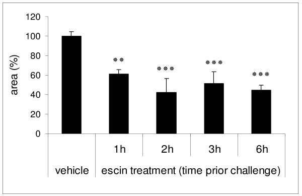
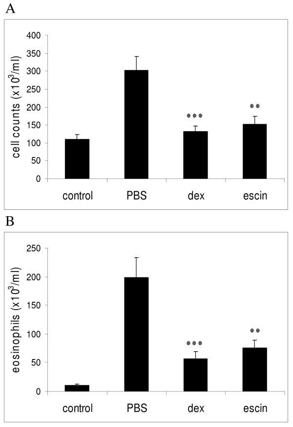
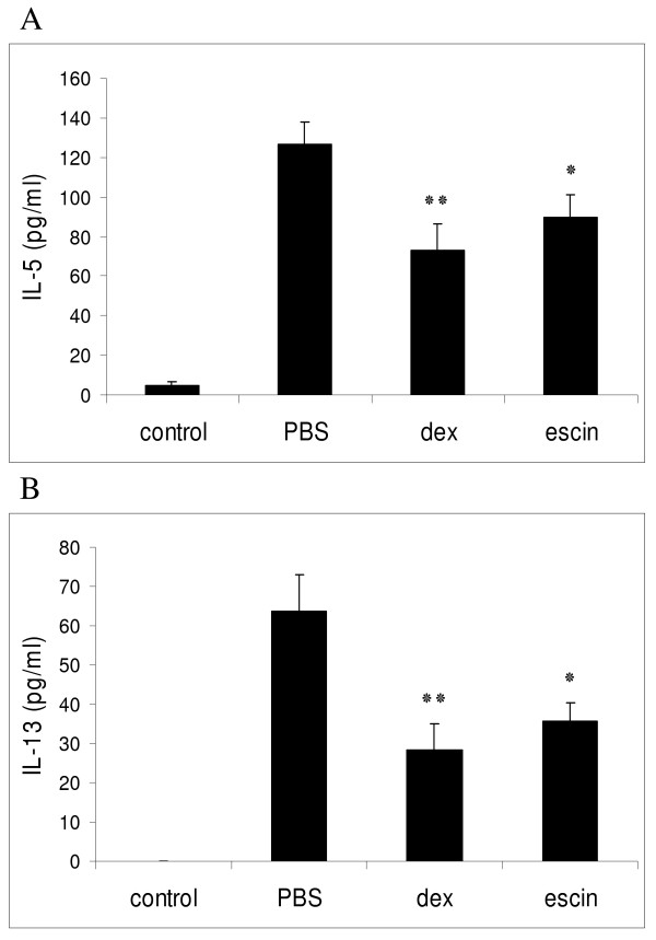
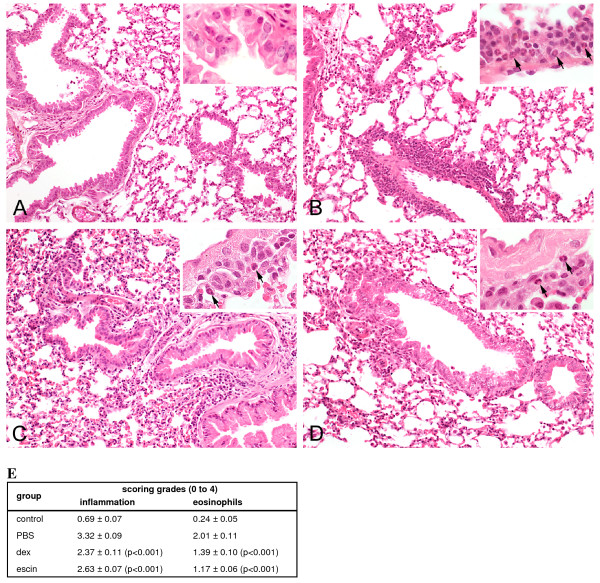
Results: In the course of a routine in vitro screening process, we identified beta-escin as a potentially anti-allergic compound. Here we tested beta-escin in two mouse models to confirm this anti-allergic effect in vivo. In a model of the early phase of allergic reactions, the murine passive cutaneous anaphylaxis model, beta-escin inhibited the effects of mast cell activation and degranulation in the skin and dose-dependently prevented the extravasation of fluids into the tissue. Beta-escin also significantly inhibited the late response after antigen challenge in a lung allergy model with ovalbumin-sensitized mice. Allergic airway inflammation was suppressed, which was exemplified by the reduction of leucocytes, eosinophils, IL-5 and IL-13 in the bronchoalveolar lavage fluid. Histopathological examinations further confirmed the reduced inflammation of the lung tissue. In both models, the inhibitory effect of beta-escin was comparable to the benchmark dexamethasone.

Conclusions: We demonstrated in two independent murine models of type I hypersensitivity that beta-escin has potent anti-allergic properties. These results and the excellent safety profile of beta-escin suggest a therapeutic potential of this compound for a novel treatment of allergic diseases.


Figures

Figure 1
Figure 1
Anti-allergic and dose-dependent efficacy of beta-escin in the murine PCA model. For PCA induction, anti-TNP IgE (2.5 μg) was injected intradermally into the dorsal skin at 6 independent spots. After 15 min, mice were challenged with intravenous injection of 50 μg DNP-BSA supplemented with 1% Evans blue dye. Blue stained skin areas were quantified from digital photographs 15 min after challenge. (A) SKH1-mice were pre-treated i.p. with vehicle (10 ml/kg, n = 21), dexamethasone (dex, 3 mg/kg, n = 5) or beta-escin (escin, 3 mg/kg, n = 12) 6 h prior application of antigen. (B) Different concentrations of beta-escin (n = 4-12) were given i.p. 6 h prior challenge. For each mouse the mean of 6 spots was calculated. Based on these values the mean ± SEM was determined for each group, normalized to the vehicle control group (equals 100%) and represented as column or point in the graphs (A) and (B), respectively. Significant differences between the treatment groups and the vehicle group are indicated with: * p < 0.05 and *** p < 0.001.
Figure 2
Figure 2
Effects of beta-escin at different time points of pre-treatment in the murine PCA model. SKH1-mice were injected with beta-escin (3 mg/kg) at different time points prior PCA induction as described in Fig. 1. For each mouse the mean of 6 spots was calculated. Based on these values the mean ± SEM was determined for 4 - 12 (escin) or 21 (vehicle) mice per treatment group and normalized to the vehicle control group. Significant differences between the treatment groups and the vehicle group are indicated with: ** p < 0.01 and *** p < 0.001.
Figure 3
Figure 3
Beta-escin reduces total cells and eosinophil recruitment in an OVA-induced model of allergic airway inflammation. BALB/c mice were sensitized with OVA on days 0, 14 and 21. The aerosol challenges with OVA or PBS were performed twice a day on days 28 and 29. 40 h after the last aerosol challenge, the mice were sacrificed for the analyses of the airway inflammation of the lungs. Groups were treated as follows: control group = PBS-challenged, treated with PBS; PBS group = OVA-challenged, treated with PBS; dex group = OVA-challenged, treated with dexamethasone (1 mg/kg, once a day) and escin group = OVA-challenged, treated with beta-escin (3 mg/kg, twice a day). Groups were compared with respect to the total cell counts (A) and the number of eosinophils (B) in the BALFs. Each bar represents the mean of 13 - 21 mice from 4 independent, but identical experiments ± SEM out of a series of similar experiments. Significant differences of the dex and escin groups to the PBS group are indicated with: ** p < 0.01 and *** p < 0.001.
Figure 4
Figure 4
Beta-escin reduces cytokine production in an OVA-induced model of allergic airway inflammation. BAL fluid was collected 15 h after the last OVA-challenge from sensitized BALB/c mice of the 4 different treatment groups as described in Fig. 3. (A) IL-5 and (B) IL-13 levels in the BALF of individual mice from each group were determined by ELISA. Each bar represents the mean of 11 - 16 mice from 3 independent, but identical experiments ± SEM out of a series of similar experiments. Significant differences of the dex and escin groups to the PBS group are indicated with: * p < 0.05 and ** p < 0.01.
Figure 5
Figure 5
Beta-escin reduces inflammation and eosinophilia in lungs of mice from the allergic airway inflammation model. Representative photomicrographs in a 20 and 60 times magnification (inset) of H&E stained lung sections from 40 h after the last challenge show the lung parenchyma and the peribronchial and perivascular infiltration of inflammatory cells from (A) PBS-challenged mice treated with PBS (negative control); (B) OVA-challenged mice treated with PBS (= positive control); (C) OVA-challenged mice treated with dexamethasone (1 mg/kg, once a day) and (D) OVA-challenged mice treated with beta-escin (3 mg/kg, twice a day). The arrows indicate representative eosinophils in the infiltrate. (E) Semi-quantitative analyses of peribronchial and perivascular inflammatory cells and percentage of eosinophils in lung sections were performed with a scoring system. Each value represents the mean of 13 - 21 mice from 4 independent, but identical experiments ± SEM out of a series of similar experiments. Significant differences of the dex and escin groups to the PBS group are indicated in brackets.

Similar articles

Cited by

References

    1. Devereux G. The increase in the prevalence of asthma and allergy: food for thought. Nat Rev Immunol. 2006;6:869–874. doi: 10.1038/nri1958. - DOI - PubMed
    1. Bloemen K, Verstraelen S, Van Den HR, Witters H, Nelissen I, Schoeters G. The allergic cascade: review of the most important molecules in the asthmatic lung. Immunol Lett. 2007;113:6–18. doi: 10.1016/j.imlet.2007.07.010. - DOI - PubMed
    1. Baumruker T, Prieschl E. Mast cells and their activation - from the molecular mechanism to clinical relevance. Mod Asp Immunobiol. 2001;1:259–262.
    1. Marshall JS. Mast-cell responses to pathogens. Nat Rev Immunol. 2004;4:787–799. doi: 10.1038/nri1460. - DOI - PubMed
    1. Galli SJ, Tsai M, Piliponsky AM. The development of allergic inflammation. Nature. 2008;454:445–454. doi: 10.1038/nature07204. - DOI - PMC - PubMed

Publication types

MeSH terms