Review
. 2010 Jun;9(3):117-27.

Advances in molecular imaging with ultrasound

Affiliations
Review

Advances in molecular imaging with ultrasound

Ryan Gessner et al. Mol Imaging. 2010 Jun.

Abstract

Ultrasound imaging has long demonstrated utility in the study and measurement of anatomic features and noninvasive observation of blood flow. Within the last decade, advances in molecular biology and contrast agents have allowed researchers to use ultrasound to detect changes in the expression of molecular markers on the vascular endothelium and other intravascular targets. This new technology, referred to as ultrasonic molecular imaging, is still in its infancy. However, in preclinical studies, ultrasonic molecular imaging has shown promise in assessing angiogenesis, inflammation, and thrombus. In this review, we discuss recent advances in microbubble-type contrast agent development, ultrasound technology, and signal processing strategies that have the potential to substantially improve the capabilities and utility of ultrasonic molecular imaging.


Figures

Figure 1
Figure 1
A cartoon graph illustrating how freely circulating and targeted microbubbles contribute to the overall concentration of contrast within an in vivo environment. Adapted from Christiansen JP and Lindner JR. MCA = microbubble contrast agent.
Figure 2
Figure 2
A schematic showing different microbubble shell and targeting ligand architectures. A, Multiple-targeting ligands, such as selectin and immunoglobin adhesion molecules, improve the likelihood of targeting by facilitating a rolling action along vessel walls. B, Polymeric forms of targeting ligands promote the likelihood of sustained adhesion. C, Excess lipid in shell (“wrinkly bubbles”) improves sustained microbubble retention. D, Buried-ligand architecture (“stealthy bubbles”) reduces both nonspecific binding and immunogenic reactions by veiling targeting ligands from complement proteins until transiently revealed at the desired target site.
Figure 3
Figure 3
The relationships between excitation frequency, micro-bubble radius, and resulting echo amplitude. Five-cycle excitation pulses pressure-matched to 100 kPa. Echo amplitudes for each microbubble diameter were maximized near their resonant frequencies, with the greatest achievable response increasing with increasing microbubble size. Adapted from Kaya M et al.
Figure 4
Figure 4
The relative image intensity improvement between size-isolated large targeted and control microbubbles (left) and targeted and control microbubbles with smaller unsorted polydisperse distributions (right). Injected concentrations were matched to 3 × 10⁷ bubbles/mL and imaged after freely circulating bubbles had cleared. Reproduced from Streeter JE et al.
Figure 5
Figure 5
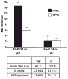
Improvement in targeting retention efficiency of wrinkled (WNKL) microbubbles (MB) compared to traditional spherical (SPHR) microbubbles. Both populations were targeted to P-selectin Rb40.34 with rat monoclonal antibodies. Data acquired using intravital microscopy in 10 different venule locations within both wild-type (WT) and P-selectin-deficient control (P−/−) mice, with n = 4 and n = 3, respectively. Adapted from Rychak JJ et al. WSS = wall shear stress.
Figure 6
Figure 6
Enzyme-linked immunosorbent assay analysis illustrating reduced immunogenic response associated with a buried-ligand architecture microbubble compared to a conventional exposed-ligand targeted bubble. Biotin ligands were used in this study. Anaphalatoxin C3a concentration measured after 30-minute in vitro incubation in human serum. “Blank” was PBS only and “control” was a distribution of bubbles bearing no ligand. Adapted from Borden MA et al.
Figure 7
Figure 7
In vitro images in a flow phantom demonstrating a method to delineate stationary from freely circulating bubbles in real time. Both freely circulating bubbles and stationary bubbles can be seen in A, although in B the signal intensity from freely circulating bubbles is suppressed with a slow-time interframe filter. Bubbles were forced to the distal wall of the vessel with radiation force pulse prior to image collection. The stationary bubble signal is indicated by the white arrow in both A and B. Field of view is 4.5 × 18 mm. Adapted from Patil AV et al.
Figure 8
Figure 8
Three-dimensional molecular imaging of angiogenesis in a rat fibrosarcoma model. The data illustrate the heterogeneity of microbubble targeting throughout the tumor volume (Streeter and colleagues, unpublished data, 2009). A, Three-dimensional rendered isosurface produced from B-mode data with manually defined regions of interest around the perimeter. B, Corresponding overlaid two-dimensional frames, as seen on an imaging system, prior to three-dimensional reconstruction. Grayscale anatomic images were collected with B-mode, whereas green overlaid contrast-only images were collected with Siemens contrast pulse sequence (Mountain View, CA).
Figure 9
Figure 9
Images and data illustrating the improvement in targeting efficiency facilitated by the application of radiation force (RF) in an angiogenic rat tumor model. A and B are three-dimensional reconstructions of two-dimensional imaging planes acquired after freely flowing bubbles cleared from the system. A, RF not administered. B, RF pulses administered for 15 seconds after bolus injection of contrast. Scale bars are 1 cm. C, Plot comparing targeted signal between acquisitions with RF application and without. A mean signal increase of 13 dB was observed across all slices in response to RF. Adapted from Gessner R et al.

Similar articles

Cited by

References

    1. Keller MW, Glasheen W, Kaul S. Albunex: a safe and effective commercially produced agent for myocardial contrast echocardiography. J Am Soc Echocardiogr. 1989;2:48–52. - PubMed
    1. Smith MD, Elion JL, McClure RR, et al. Left heart opacification with peripheral venous injection of a new saccharide echo contrast agent in dogs. J Am Coll Cardiol. 1989;13:1622–8. - PubMed
    1. Klibanov AL. Ultrasound contrast agents: development of the field and current status. In: Krause W, editor. Contrast agents II. Vol. 222. New York: Springer-Verlag; 2002. pp. 73–106.
    1. Stride E. Physical principles of microbubbles for ultrasound imaging and therapy. Cerebrovasc Dis. 2009;27 (Suppl 2):1–13. - PubMed
    1. Dayton PA, Ferrara KW. Targeted imaging using ultrasound. J Magn Reson Imaging. 2002;16:362–77. - PubMed

Publication types