Review
. 2010 Nov;29(6):500-19.
doi: 10.1016/j.preteyeres.2010.05.003. Epub 2010 May 19.

Animal models of choroidal and retinal neovascularization

Affiliations
Review

Animal models of choroidal and retinal neovascularization

Hans E Grossniklaus et al. Prog Retin Eye Res. 2010 Nov.

Abstract

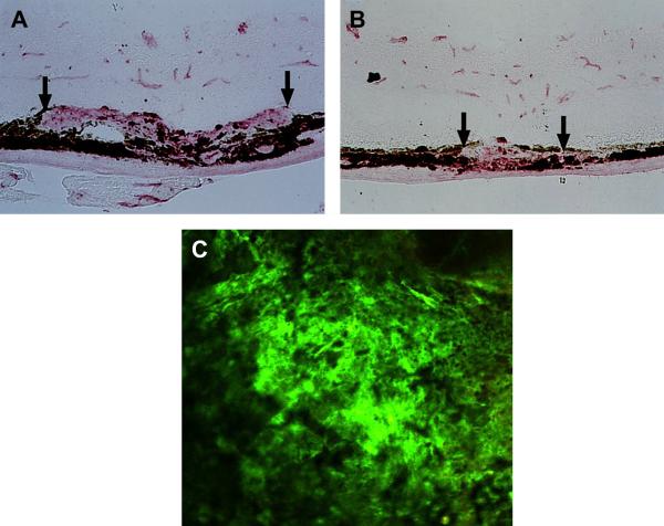
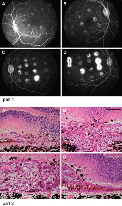
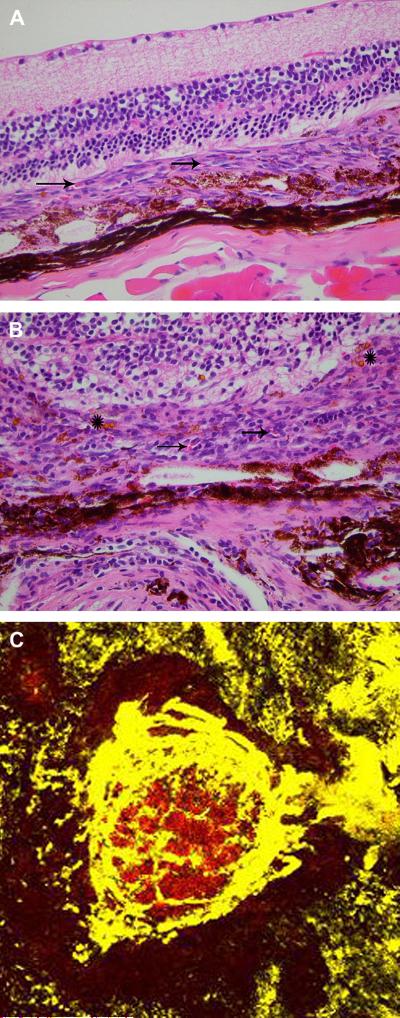
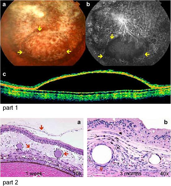
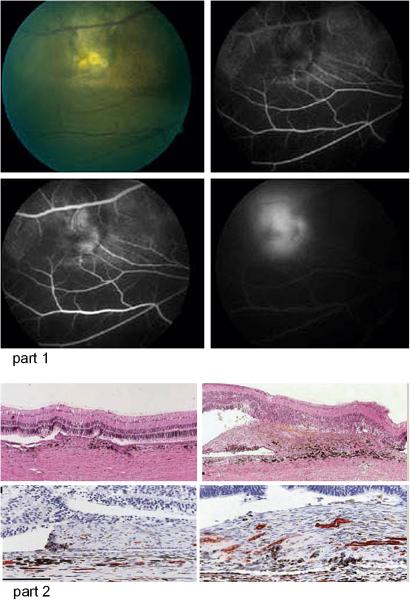
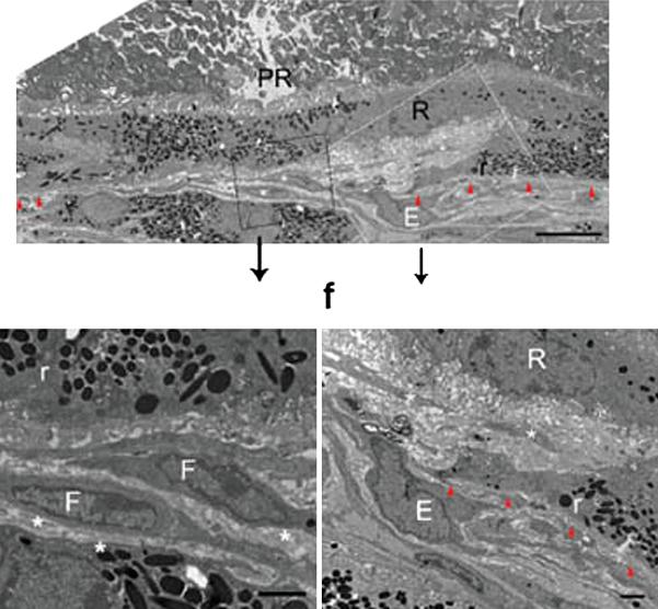
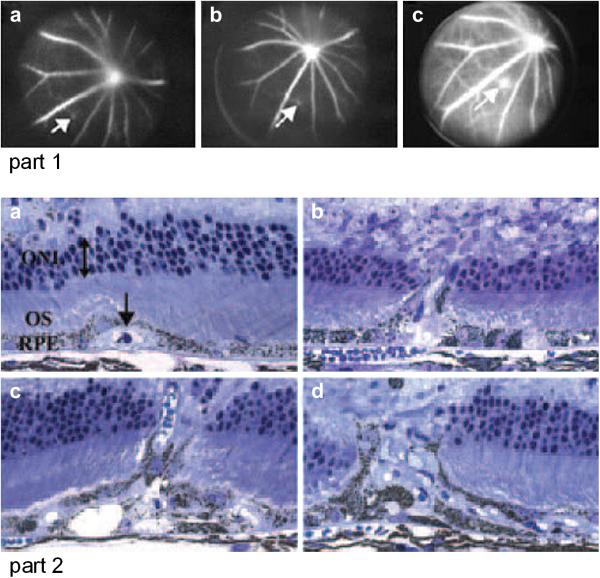
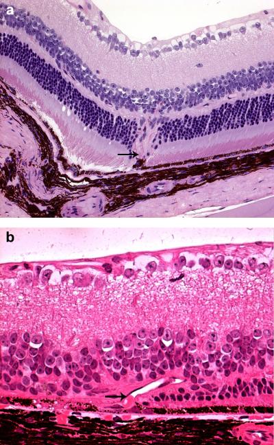
There have been numerous types of animal models of choroidal neovascularization (CNV) and retinal neovascularization (RNV). Understanding the pathobiology of CNV and RNV is important when evaluating and utilizing these models. Both CNV and RNV are dynamic processes. A break or defect in Bruchs' membrane is necessary for CNV to develop. This may be induced with a laser, mechanically via surgery, or in the setting of transgenic mice. Some of the transgenic mouse models spontaneously develop RNV and/or retinal angiomatous proliferation (RAP)-like lesions. The pathogenesis of RNV is well-known and is generally related to ischemic retinopathy. Models of oxygen-induced retinopathy (OIR) closely resemble retinopathy of prematurity (ROP). The streptozotocin (STZ) rat model develops features similar to diabetic retinopathy. This review summarizes general categories and specific examples of animal models of CNV and RNV. There are no perfect models of CNV or RNV and individual investigators are encouraged to choose the model that best suits their needs.


Figures

Figure 1
Figure 1. Mouse laser model of CNV
A. Control mouse with laser induced CNV (between arrows). B. A mouse is treated with a drug that inhibits VEGF and PDGF develops smaller CNV (between arrows) after laser induction. (From Kwak et al, 2000) C. A flat mount preparation of laser induced CNV in the mouse after perfusion with fluorescent dextran shows the horizontal extent of the CNV. (From Singh et al, 2009)
Figure 2
Figure 2. Cynomolgus monkey laser model of CNV
Part 1. Fluorescein angiography 21 days after laser induction of CNV shows partial localized (A and B) or mixed localized and diffuse CNV (C and D) Part 2. Histologic examination of enucleate eyes shows CNV between the retina and a reflected layer of RPE (A), extension of the CNV into the retina (B), and numerous vascular channels within the CNV which also contains pigmented macrophages (C and D). (From Criswell et al, 2004)
Figure 3
Figure 3. Mouse surgical model of CNV
A. After subretinal injection of RPE and polystyrene beads, CNV is located between the retina and a reflected layer of RPE. Vascular channels are indicated by arrows. B. There are vascular channels (arrows), mononuclear inflammatory cells, and beads (asterisks) located within the CNV. C. A fluorescent labeled dextran flatmount preparation shows the extent of the CNV. (From Schmack et al, 2009)
Figure 4
Figure 4. Rabbit surgical model of CNV
Part 1. A color fundus photograph (a), fluorescein-dextran angiogram (b) and OCT show that there is a separation of the neurosensory from the underlying Bruchs membrane and choroid. Part 2. CNV is present in the subretinal space 1 week (a) and 3 months (b) after subretinal injection of 100 ng FGF-2/100 ng LPS. There are inflammatory cells present (arrows) in the retina, CNV, and surrounding Heparin-sepharose beads at the injection area in the 1 week old CNV. There are giant cells (open arrow) in the 3 month old CNV. (From Ni et al, 2005)
Figure 5
Figure 5. Pig surgical model of CNV
Part 1. A fundus photograph (upper left) and fluorsecein angiogram early (upper right), middle (bottom left) and late (bottom right) phases show a yellow, early hyperfluorescent with late leakage CNV lesion in the pig model with perforation of Bruchs membrane only. Part 2. Histology and immunohistochemical staining for CD34 that shows vascular channels shows tha the CNV lesion is smaller and has fewer vascular channels after RPE removal/Bruchs perforation (upper and lower left) compared with Bruchs membrane perforation alone (upper and lower right). (From Lassota et al 2007)
Figure 6
Figure 6
Mouse CNV in Ccr2/CCl2 transgenic model Electron micrographs of CNV shows endothelial cells (E) and fibrocytes (F) from the choroid breaking through Bruchs membrane and extending into the sub-RPE (R) space. Photoreceptors (PR) are present internal to the RPE. (From Ambati et al, 2003)
Figure 7
Figure 7. Mouse ApoE overexpression transgenic model of CNV
Part 1. Early (a), mid (b) and late-phase (c) fluorescein angiogram demonstrates area of hyperfluorescence and leakage indicative of CNV. Part 2. Histology showing CNV under RPE (a), extending from the sub-RPE space into the retina (b and c), and extensive CNV (d). (From Malek et al, 2005)
Figure 8
Figure 8. Mouse Ccl2Cx3cr1 transgenic mouse model of subretinal neovascualrization
There is neovascularization in the deep retina with unvolvement of the underlying RPE (A). These vascular channels (arrow) are present under the photoreceptors and appear to arise from the area of the RPE (arrow) (B). Courtesy of Chi Chao Chan MD
Figure 9
Figure 9. Schematic diagram of the cascade of events that have been implicated in the development of retinal neovascularization
Abbreviations: HIF-1, hypoxia-inducible factor 1; IGF-1, insulin-like growth factor 1; PDGF-BB, platelet-derived factor B chain homodimer; TGF-βs, members of the transforming growth factor beta super family; VEGF, vascular endothelial growth factor; VEGFR1, VEGF receptor 1. (From Campochiaro, 2000)
Figure 10
Figure 10. Time course of retinal vascular response to hyperoxia in fluorescein-dextran perfused flat mounts
Left: normal P7 retina before hyperoxia exposure. Note area of nonperfusion in periphery where blood vessels have not yet developed. Right: P14 retina after 5 days of hyperoxia exposure, followed by 2 days in room air. Note increased perfusion in the periphery and dilation and tortuosity of radial vessels and persistent absence of central perfusion. (Adapted from Smith et al,1994)
Figure 11
Figure 11. Retinal flat mount of rat OIR model
Peripheral neovascularization is seen in the rat retina. (From Madan and Penn, 2003)
Figure 12
Figure 12
Characterization of different transgenic mouse lines by color fundus photography, fluorescein fundus angiography, fluorescein micrographs of flat mounted, fluorescein labeled dextran perfused eyes, and light micrographs of hematoxylin and eosin stained paraffin sections. (From Lai et al, 2005)

Similar articles

Cited by

References

    1. Aguilar E, Dorrell MI, Friedlander D, Jacobson RA, Johnson A, Marchetti V, Moreno SK, Ritter MR, Friedlander M. Ocular models of angiogenesis. Methods Enzymol. 2008;444:115–158. - PubMed
    1. Albert DM, Neekhar A, Wang S, Darjatmoko SR, Sorenson CM, Subielzig RR, Sheibani N. Development of choroidal neovascularizaiton in rats with advanced intense cyclic light-induced retinal degeneration. Arch Ophthalmol. 2010;128:212–222. - PMC - PubMed
    1. Ambati J, Anand A, Fernandez S, et al. An animal model of age-related macular degeneration in senescent Ccl-2- or Ccr-2- deficient mice. Nat Med. 2003;11:1290–1397. - PubMed
    1. Amin R, Pucklin JE, et al. Growth factor localization in choroidal noevascular membranes of age-related macular degeneration. Inv Ophthalmol Vis Sci. 1994;35:3178–3188. - PubMed
    1. Antoszyk AN, Gottlieb JL, Machemer R, Hatchell DL. The effects of intravitreal triamcinolone acetonide on experimental pre-retinal neovascularization. Graefes Arch Clin Exp Ophthalmol. 1993;231:34–40. - PubMed

Publication types

MeSH terms

LinkOut - more resources