

Carbamylation-dependent activation of T cells: a novel mechanism in the pathogenesis of autoimmune arthritis
- PMID: 20488785
- PMCID: PMC2925534
- DOI: 10.4049/jimmunol.1000075
Carbamylation-dependent activation of T cells: a novel mechanism in the pathogenesis of autoimmune arthritis
Abstract
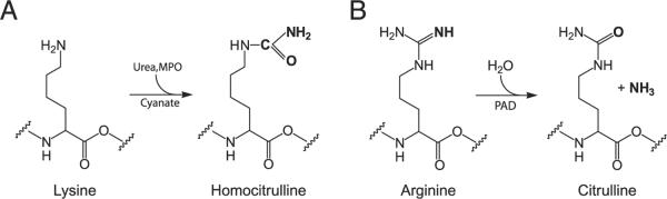
The posttranslational modification of proteins has the potential to generate neoepitopes that may subsequently trigger immune responses. The carbamylation of lysine residues to form homocitrulline may be a key mechanism triggering inflammatory responses. We evaluated the role of carbamylation in triggering immune responses and report a new role for this process in the induction of arthritis. Immunization of mice with homocitrulline-containing peptides induced chemotaxis, T cell activation, and Ab production. The mice also developed erosive arthritis following intra-articular injection of peptides derived from homocitrulline and citrulline. Adoptive transfer of T and B cells from homocitrulline-immunized mice into normal recipients induced arthritis, whereas systemic injection of homocitrulline-specific Abs or intra-articular injection of homocitrulline-Ab/citrulline-peptide mixture did not. Thus, the T cell response to homocitrulline-derived peptides, as well as the subsequent production of anti-homocitrulline Abs, is critical for the induction of autoimmune reactions against citrulline-derived peptides and provides a novel mechanism for the pathogenesis of arthritis.
Figures


Similar articles
-
Ureido group-specific antibodies are induced in rabbits immunized with citrulline- or homocitrulline-containing antigens.Autoimmunity. 2016 Nov;49(7):459-465. doi: 10.3109/08916934.2016.1171853. Epub 2016 Apr 21. Autoimmunity. 2016. PMID: 27098309
-
Immune responses to peptides containing homocitrulline or citrulline in the DR4-transgenic mouse model of rheumatoid arthritis.J Autoimmun. 2018 May;89:75-81. doi: 10.1016/j.jaut.2017.12.002. Epub 2017 Dec 11. J Autoimmun. 2018. PMID: 29242008
-
Rheumatoid arthritis-associated rheumatoid factors post-COVID-19.Front Immunol. 2025 Feb 13;16:1553540. doi: 10.3389/fimmu.2025.1553540. eCollection 2025. Front Immunol. 2025. PMID: 40018049 Free PMC article.
-
Pitfalls in the detection of citrullination and carbamylation.Autoimmun Rev. 2018 Feb;17(2):136-141. doi: 10.1016/j.autrev.2017.11.017. Epub 2017 Dec 2. Autoimmun Rev. 2018. PMID: 29203292 Review.
-
Autoimmunity in rheumatoid arthritis: different antigens--common principles.Ann Rheum Dis. 2013 Apr;72 Suppl 2:ii132-6. doi: 10.1136/annrheumdis-2012-202349. Epub 2012 Dec 19. Ann Rheum Dis. 2013. PMID: 23253931 Review.
Cited by
-
Antibodies to carbamylated α-enolase epitopes in rheumatoid arthritis also bind citrullinated epitopes and are largely indistinct from anti-citrullinated protein antibodies.Arthritis Res Ther. 2016 May 4;18(1):96. doi: 10.1186/s13075-016-1001-6. Arthritis Res Ther. 2016. PMID: 27145822 Free PMC article.
-
Complementary Effects of Carbamylated and Citrullinated LL37 in Autoimmunity and Inflammation in Systemic Lupus Erythematosus.Int J Mol Sci. 2021 Feb 6;22(4):1650. doi: 10.3390/ijms22041650. Int J Mol Sci. 2021. PMID: 33562078 Free PMC article.
-
Immunopathogenesis of Rheumatoid Arthritis.Immunity. 2017 Feb 21;46(2):183-196. doi: 10.1016/j.immuni.2017.02.006. Immunity. 2017. PMID: 28228278 Free PMC article. Review.
-
The Therapeutic Potential of Phytochemicals Unlocks New Avenues in the Management of Rheumatoid Arthritis.Int J Mol Sci. 2025 Jul 16;26(14):6813. doi: 10.3390/ijms26146813. Int J Mol Sci. 2025. PMID: 40725063 Free PMC article. Review.
-
Localisation of citrullinated and carbamylated proteins in inflamed gingival tissues from rheumatoid arthritis patients.Clin Oral Investig. 2021 Mar;25(3):1441-1450. doi: 10.1007/s00784-020-03452-9. Epub 2020 Jul 12. Clin Oral Investig. 2021. PMID: 32656595
References
-
- Anderton SM. Post-translational modifications of self antigens: implications for autoimmunity. Curr. Opin. Immunol. 2004;16:753–758. - PubMed
-
- Mann E, McDermott MJ, Goldman J, Chiesa R, Spector A. Phosphorylation of alpha-crystallin B in Alexander's disease brain. FEBS Lett. 1991;294:133–136. - PubMed
-
- Doyle HA, Mamula MJ. Posttranslational modifications of self-antigens. Ann. N. Y. Acad. Sci. 2005;1050:1–9. - PubMed
-
- Zamvil SS, Mitchell DJ, Moore AC, Kitamura K, Steinman L, Rothbard JB. T-cell epitope of the autoantigen myelin basic protein that induces encephalomyelitis. Nature. 1986;324:258–260. - PubMed
Publication types
MeSH terms
Substances
Grants and funding
LinkOut - more resources
Full Text Sources
Other Literature Sources
Medical
