. 2010 Oct;31(9):1596-601.
doi: 10.3174/ajnr.A2123. Epub 2010 May 20.

Septopreoptic holoprosencephaly: a mild subtype associated with midline craniofacial anomalies

Affiliations

Septopreoptic holoprosencephaly: a mild subtype associated with midline craniofacial anomalies

J S Hahn et al. AJNR Am J Neuroradiol. 2010 Oct.

Abstract

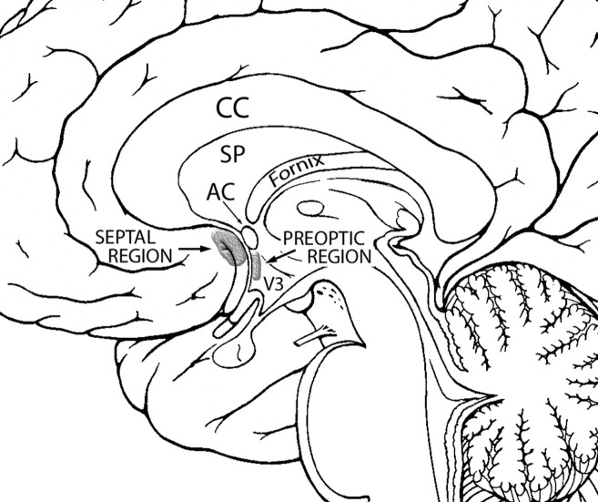
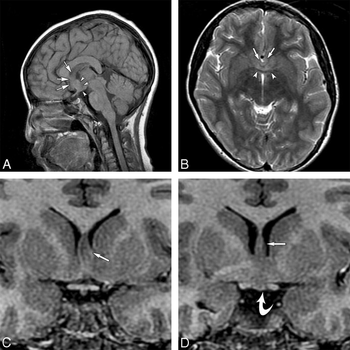
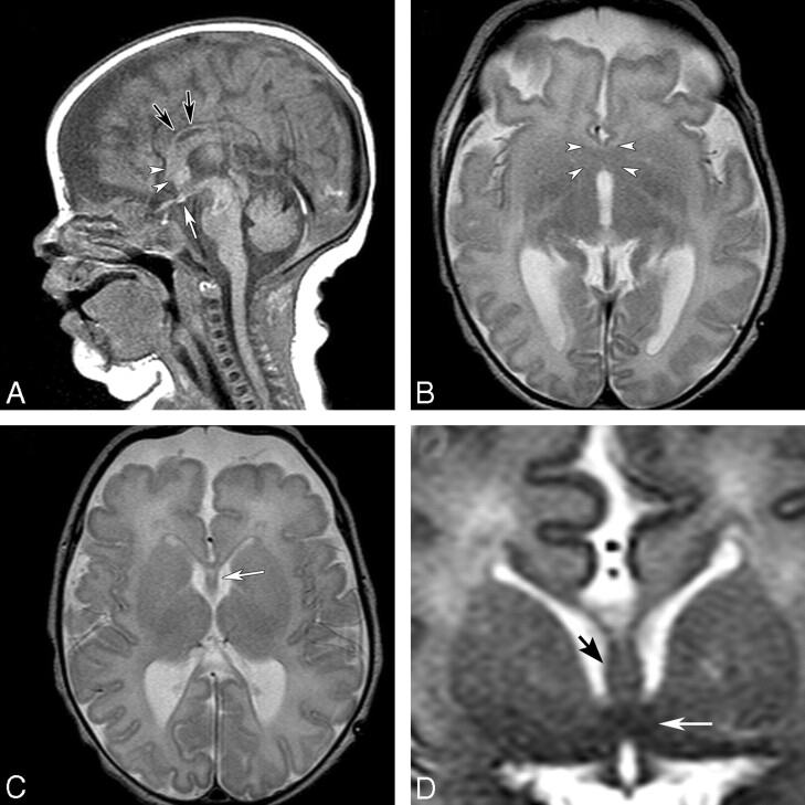
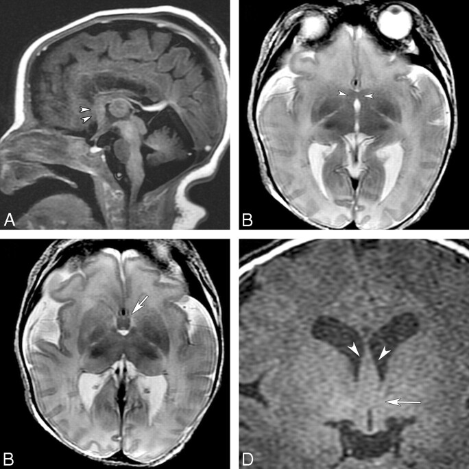
HPE is a congenital brain malformation characterized by failure of the prosencephalon to divide into 2 hemispheres. We have identified 7 patients who have a mild subtype of HPE in which the midline fusion was restricted to the septal region or preoptic region of the telencephalon. This subtype, which we call septopreoptic HPE, falls in the spectrum of lobar HPE, but lacks significant frontal neocortical fusion seen in lobar HPE. Other imaging characteristics include thickened or dysplastic fornix, absent or hypoplastic anterior CC, and single unpaired ACA. The SP was fully formed in 4, partially formed in 2, and absent in 1. Mild midline craniofacial malformation, such as SMMCI and CNPAS were found in 86% and 71%, respectively. Patients outside of infancy often manifested language delay, learning disabilities, or behavioral disturbances, while motor function was relatively spared.


Figures

Fig 1.
Fig 1.
Diagram of the midsagittal view of the brain displaying the locations of the septal and preoptic regions. Modified from Martin J.
Fig 2.
Fig 2.
MR imaging of a 10-year-old boy with learning disabilities, a SMMCI, CNPAS, precocious puberty, and other endocrinopathies. A, T1-weighted midsagittal image shows hypoplasia of the rostrum of the CC and a rectangular area of abnormality in the subcallosal region, anterior to the hypothalamic region (arrowheads). The dysplastic-appearing fornix is anterior to this region (arrows). B, Axial T2-weighted image shows well-developed anterior and posterior interhemispheric fissures and an azygous ACA flow void in the anterior interhemispheric fissure. There is an area of midline fusion just anterior to the AC, which appears as a dark bowlike band (arrowheads). Further anterior to this region are the dysplastic fornix (arrows) and cortical gray matter that is continuous in the middle. C, Coronal SPGR image anterior to the AC shows an area of fusion of the septal region (arrow). D, Coronal SPGR image at the level to the AC shows dysplastic thickened fornices (arrow) traveling below the SP and inferiorly an area of midline fusion in the preoptic region and basal structures (curved arrow).
Fig 3.
Fig 3.
MR imaging of an 8-day-old term neonate with atrial and ventricular septal defects and SMMCI. A, Sagittal T1-weighted image shows a thin CC (black arrows), a thickened fornix, and dysplastic subcallosal areas (arrowheads). A small ectopic pituitary gland is noted near the chiasm (white arrow). B, Axial T2-weighted image shows an area of fusion in the septal and preoptic regions (arrowheads). C, An azygous ACA is present in the anterior IHF. Axial T2-weighted image slightly superior shows the presence of the SP (white arrow) and a thin genu of the CC. D, Coronal T2-weighted image at the level of the AC shows fusion of the midline region (white arrow) below the fornices (black arrow).
Fig 4.
Fig 4.
MR imaging of a 6-day-old term neonate initially diagnosed as having choanal atresia, vertebral anomalies, and coactation of aorta. A, Sagittal FSPGR image shows an area of fusion in the subcallosal region (arrowheads). B, Axial T2-weighted image shows the abnormal fusion in the same region (arrowheads). An azygous ACA is present in the anterior IHF. C, Axial T2-weighted image slightly superior shows thickened fornices (arrow) and partial fusion of the thalami. D, Coronal FSPGR image shows thickened fornices (arrowheads) and an area of fusion in the preoptic area (arrow).

References

    1. Golden JA. Towards a greater understanding of the pathogenesis of holoprosencephaly. Brain Dev 1999; 21: 513– 21 - PubMed
    1. DeMyer W. Holoprosencephaly (cyclopia-arrhinencephaly). In: Vinken PJ, Bruyn GW, Klawans HL.eds. Handbook of Clinical Neurology. 6th ed. Amsterdam, the Netherlands: Elsevier Science Publishers; 1987: 225– 44
    1. Lewis AJ, Simon EM, Barkovich AJ, et al. Middle interhemispheric variant of holoprosencephaly: a distinct cliniconeuroradiologic subtype. Neurology 2002; 59: 1860– 65 - PubMed
    1. Simon EM, Hevner RF, Pinter JD, et al. The middle interhemispheric variant of holoprosencephaly. AJNR Am J Neuroradiol 2002; 23: 151– 55 - PMC - PubMed
    1. Simon EM, Hevner R, Pinter JD, et al. Assessment of the deep gray nuclei in holoprosencephaly. AJNR Am J Neuroradiol 2000; 21: 1955– 61 - PMC - PubMed

Publication types