. 2010 Aug;116(2):577-89.
doi: 10.1093/toxsci/kfq148. Epub 2010 May 20.

Silver nanoparticles disrupt GDNF/Fyn kinase signaling in spermatogonial stem cells

Affiliations

Silver nanoparticles disrupt GDNF/Fyn kinase signaling in spermatogonial stem cells

Laura K Braydich-Stolle et al. Toxicol Sci. 2010 Aug.

Abstract

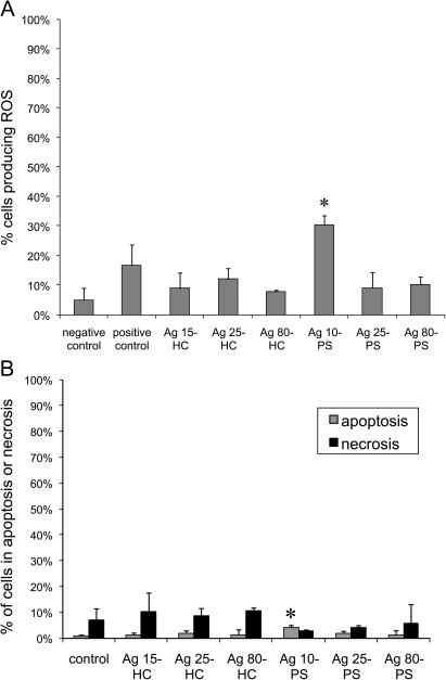
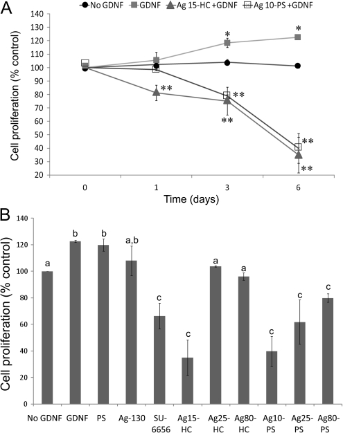
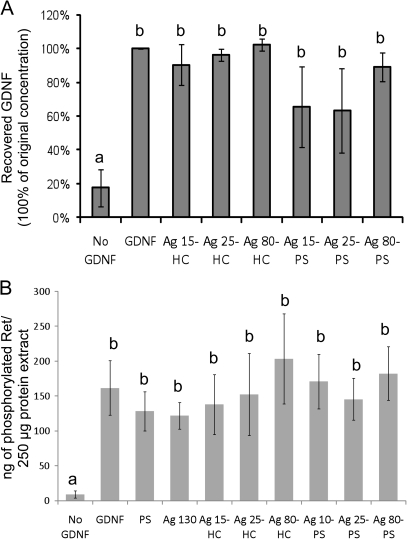
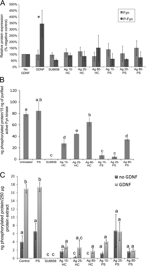
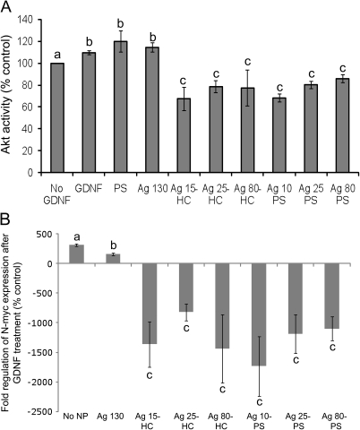
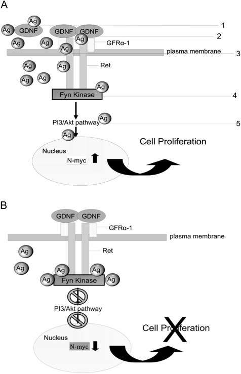
Silver nanoparticles (Ag-NPs) are being utilized in an increasing number of fields and are components of antibacterial coatings, antistatic materials, superconductors, and biosensors. A number of reports have now described the toxic effects of silver nanoparticles on somatic cells; however, no study has examined their effects on the germ line at the molecular level. Spermatogenesis is a complex biological process that is particularly sensitive to environmental insults. Many chemicals, including ultrafine particles, have a negative effect on the germ line, either by directly affecting the germ cells or by indirectly acting on the somatic cells of the testis. In the present study, we have assessed the impact of different doses of Ag-NPs, as well as their size and biocompatible coating, on the proliferation of mouse spermatogonial stem cells (SSCs), which are at the origin of the germ line in the adult testis. At concentrations >OR= 10 microg/ml, Ag-NPs induced a significant decline in SSCs proliferation, which was also dependent on their size and coating. At the concentration of 10 microg/ml, reactive oxygen species production and/or apoptosis did not seem to play a major role; therefore, we explored other mechanisms to explain the decrease in cell proliferation. Because glial cell line-derived neurotrophic factor (GDNF) is vital for SSC self-renewal in vitro and in vivo, we evaluated the effects of Ag-NPs on GDNF-mediated signaling in these cells. Although the nanoparticles did not reduce GDNF binding or Ret receptor activity, our data revealed that already at a concentration of 10 microg/ml, silver nanoparticles specifically interact with Fyn kinase downstream of Ret and impair SSC proliferation in vitro. In addition, we demonstrated that the particle coating was degraded upon interaction with the intracellular microenvironment, reducing biocompatibility.


Figures

FIG. 1.
FIG. 1.
Surface binding of coated silver nanoparticles by SSCs. Scanning electron micrographs of C18-4 cells treated with silver nanoparticles and taken at ×1300 magnification. The largest particles were rather found in aggregates, whereas the smaller particles tend to scatter at the cell surface. A: control cells, B: cells treated with polysaccharide, C: Ag 130 particles treatment, D: Ag 80-HC nanoparticles treatment, E: Ag 25-HC nanoparticles treatment, F: Ag 15-HC nanoparticles treatment, G: Ag 80-PS nanoparticles treatment, H: Ag 25-PS nanoparticles treatment, and I: Ag 10-PS nanoparticles treatment. N, nucleus; Cy, cytoplasm. Arrows indicate nanoparticles and aggregates. Arrowheads indicate internalized nanoparticles.
FIG. 2.
FIG. 2.
Internalization and localization of coated silver nanoparticles in SSCs. Transmission electron micrographs of C18-4 cells treated with 10 μg/ml silver nanoparticles (scale bar = 1 μm) confirming that the particles are internalized and can be sequestered in endosomes. A: Ag 15-HC nanoparticles treatment, B: Ag 25-HC nanoparticles treatment, C: Ag 80-HC nanoparticle treatment, D: Ag 10-PS nanoparticles treatment, E: Ag 25-PS nanoparticles treatment, and F: Ag 80-PS nanoparticles treatment. Arrows indicate nanoparticles and aggregates.
FIG. 3.
FIG. 3.
Lysosomal and cytosolic localization of coated silver nanoparticles in C18-4 cells. The nanoparticles were excited at a wavelength of 375 nm and pseudocolored green, whereas the lysosomes were immunostained and excited at a wavelength of 647 nm giving the lysosomes a red color. Any nanoparticle localization within lysosomes appeared yellow. Nuclei were stained with standard DAPI. A: control cells without nanoparticles (red, arrows, indicate lysosomes). B: cells that were cultured with Ag25-HC (10 μg/ml). At this concentration, many nanoparticles were localized in lysosomes (yellow, arrowheads); however, others appear dispersed in the cytosol (green, arrows), indicating the potential for interactions with other organelles and cytosolic proteins. C: cells that were cultured with Ag25-HC (50 μg/ml). At higher concentrations, nanoparticles are mostly found in the lysosomes (yellow, arrowheads).
FIG. 4.
FIG. 4.
Viability of C18-4 cells treated with different concentrations of coated silver nanoparticles. C18-4 cells were incubated for 24 h with HC- and PS-coated nanoparticles of different sizes and concentrations, and cell viability was assessed by using the MTS assay. A: Effect of HC-coated nanoparticles. This graph shows that nanoparticles with a smaller size (Ag 15-HC and Ag 25-HC) significantly inhibit cell viability (asterisk, p < 0.005) at concentrations ≥ 10 μg/ml in comparison with the control. B: Effect of PS-coated nanoparticles. This graph shows an inhibition of cell viability at concentrations ≥ 10 μg/ml (Ag 25-PS and Ag 80-PS nanoparticles) and concentrations ≥ 25 μg/ml (Ag 15-PS nanoparticles) in comparison with the control (asterisk, p < 0.05).
FIG. 5.
FIG. 5.
Effect of coated silver nanoparticles on cellular stress responses. A: ROS production in the C18-4 cells. Following a 48-h incubation with 10 μg/ml HC- and PS-coated nanoparticles, ROS production was assessed by using the Image IT Live Green ROS Detection Kit. Significant ROS production could be detected only with the Ag 10-PS nanoparticles (asterisk, p < 0.005). B: effect of silver nanoparticles on cellular apoptosis. Apoptosis in the C18-4 cells was evaluated following a 48-h incubation with 10 μg/ml HC- and PS-coated nanoparticles using the Vybrant apoptosis assay. A small (5%) but significant number of cells undergoing apoptosis could be detected but only with the Ag 10-PS nanoparticles (asterisk, p < 0.005). Hydrocarbon-coated silver nanoparticles induced an increase in necrosis in comparison with the PS-coated nanoparticles, but the differences compared with the control were not significant.
FIG. 6.
FIG. 6.
Proliferation of C18-4 cells treated with coated silver nanoparticles. A: growth curve of C18-4 cells continuously exposed to 10 μg Ag 15-HC and Ag 10-PS silver nanoparticles and treated daily with GDNF during 6 days. The Ag 10-PS nanoparticles are initially more biocompatible, but over time, this is lost and there is no difference in growth between the two treatments. Furthermore, GDNF is unable to significantly promote proliferation when compared with the controls with no particles or GDNF (*significant increase in cell number compared with the control and **significant decrease in cell number compared with the control, p < 0.05). B: viability of C18-4 cells after 6 days of continuous exposure to silver nanoparticles and daily treatment with GDNF. GDNF was able to significantly promote cell growth when the nanoparticles were not present, but the addition of silver nanoparticles (regardless of size or coating) inhibited this effect (different letters indicate statistically significant differences, p < 0.05). PS, polysaccharide alone; SU6656, SFK inhibitor.
FIG. 7.
FIG. 7.
Effect of coated silver nanoparticles on the extracellular components of the GDNF signaling pathway. A: GDNF (100 ng/ml) was incubated for 48 h in presence of 10 μg/ml silver nanoparticles in tissue culture media. Thereafter, the particles were precipitated by centrifugation and the levels of remaining GDNF measured by ELISA. The presence of silver nanoparticles did not significantly alter the levels of free GDNF in the culture media, indicating that receptor-ligand interactions can occur. B: When C18-4 cells were treated with 10 μg/ml silver nanoparticles, GDNF was still able to promote phosphorylation of the Ret receptor, regardless of particle size or coating. Therefore, coated silver nanoparticles do not impair receptor-ligand binding and receptor activation. In both graphs, different letters indicate statistically significant differences, p < 0.05.
FIG. 8.
FIG. 8.
Fyn kinase phosphorylation and activation in the presence of coated silver nanoparticles. A: phosphorylation of Fyn kinase. The C18-4 cells were treated with 10 μg/ml silver nanoparticles for 24 h. After adding GDNF for 4 h (100 ng/ml), Fyn kinase was immunoprecipitated and its phosphorylation assessed by Western blotting. For quantitative analysis, the band intensities were evaluated with Image J Analysis software (National Institutes of Health). Western blots are presented in Supplementary figure 4. GDNF significantly increased Fyn phosphorylation in comparison with the control sample without GDNF (asterisk, p < 0.05), whereas GDNF-induced phosphorylation was inhibited by the silver nanoparticles. B: activity of purified Fyn kinase. The ex vivo influence of silver nanoparticles on the phosphorylating activity of a purified Fyn kinase was measured using a tyrosine kinase assay. The purified Fyn kinase showed a significant size-dependent decrease of activity (p < 0.05). Interestingly, the Ag-PS nanoparticles inhibited Fyn activity more than the Ag-HC nanoparticles, indicating that nanoparticle coatings have an effect on protein activity. C: activity of cellular Fyn kinase. C18-4 cells were treated with 10 μg/ml silver nanoparticles for 24 h and then treated with 100 ng/ml GDNF for 4 h. After Fyn immunoprecipitation, its activity was measured using a tyrosine kinase assay. A significant decline in Fyn kinase activity in C18-4 cells was observed in presence of 10 μg/ml nanoparticles (p < 0.05). However, this decline was not size or coating dependent. In graphs B and C, different letters indicate statistically significant differences, p < 0.05.
FIG. 9.
FIG. 9.
Effect of coated silver nanoparticles in C18-4 cells downstream of Fyn. A: Effect on Akt activity: C18-4 cells were treated with 10 μg/ml silver nanoparticles for 24 h and then with 100 ng/ml GDNF for 4 h. Akt kinase was immunoprecipitated and its activity measured with an Akt activity assay. Akt activity significantly declined in C18-4 cells treated with 10 μg/ml silver nanoparticles, and this effect was independent of size and coating. B: Effect of N-myc expression: Using qPCR, a significant decline in N-myc expression was demonstrated for C18-4 cells treated with silver nanoparticles that corresponded to the Akt activity (A), and this effect occurred independent of nanoparticle size and coating. In both graphs, different letters indicate statistically significant differences, p < 0.05.
FIG. 10.
FIG. 10.
Illustration of GDNF signaling disruption by coated silver nanoparticles. A: potential targets for disruption of GDNF signaling in C18-4 cells that inhibit proliferation. (1) Nanoparticles bind to GDNF and prevent receptor binding, (2) nanoparticles bind to receptors and prevent GDNF from binding, (3) nanoparticles interact with the Ret receptor and prevent activation of the receptor and downstream elements, (4) nanoparticles bind to Fyn kinase and prevent activation of Fyn and downstream elements, and (5) nanoparticles interact with proteins in the PI3/Akt pathway. B: model of C18-4 cell growth disruption by silver nanoparticles. The silver nanoparticles promoted a decrease in Fyn activity, which disrupted Akt activity and promoted a decline in N-myc expression, leading ultimately to a decline in cell proliferation. GDNF binding and Ret activation are not affected.

Similar articles

Cited by

References

    1. Ahamed M, Karns M, Goodson M, Rowe J, Hussain S, Schlager J, Hong Y. DNA damage response to different surface chemistry of silver nanoparticles in mammalian cells. Toxicol. Appl. Pharmacol. 2008;233:404–410. - PubMed
    1. Ahamed M, Posgai R, Gorey T, Nielsen M, Hussain S, Rowe J. Silver nanoparticles induced heat shock protein 70, oxidative stress and apoptosis in Drosophila melanogaster. Toxicol. Appl. Pharmacol. 2010;242:263–269. - PubMed
    1. Anway MD, Skinner M. Epigenetic programming of the germ line: effects of endocrine disruptors on the development of transgenerational disease. Reprod. Biomed. Online. 2008;16:23–25. - PMC - PubMed
    1. Asharani P, Hande M, Valiyaveettil S. Anti-proliferative activity of silver nanoparticles. BMC Cell Biol. 2009;10:65. - PMC - PubMed
    1. Asharani P, Low Kah Mun G, Hande M, Valiyaveettil S. Cytotoxicity and genotoxicity of silver nanoparticles in human cells. ACS Nano. 2009;3:279–290. - PubMed

Publication types

Substances