. 2010 Aug;21(8):1317-25.
doi: 10.1681/ASN.2010020134. Epub 2010 May 20.

miR-192 mediates TGF-beta/Smad3-driven renal fibrosis

Affiliations

miR-192 mediates TGF-beta/Smad3-driven renal fibrosis

Arthur C K Chung et al. J Am Soc Nephrol. 2010 Aug.

Abstract

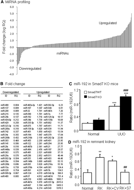
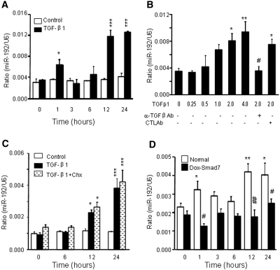
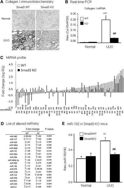
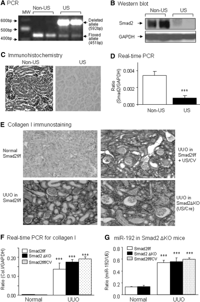
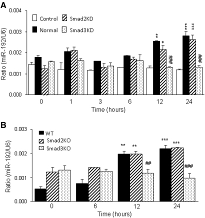
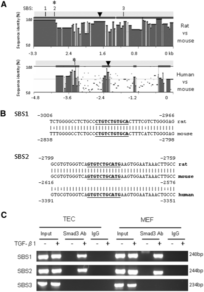
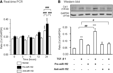
TGF-beta/Smad3 promotes renal fibrosis, but the mechanisms that regulate profibrotic genes remain unclear. We hypothesized that miR-192, a microRNA expressed in the kidney may mediate renal fibrosis in a Smad3-dependent manner. Microarray and real-time PCR demonstrated a tight association between upregulation of miR-192 in the fibrotic kidney and activation of TGF-beta/Smad signaling. Deletion of Smad7 promoted miR-192 expression and enhanced Smad signaling and fibrosis in obstructive kidney disease. In contrast, overexpression of Smad7 to block TGF-beta/Smad signaling inhibited miR-192 expression and renal fibrosis in the rat 5/6 nephrectomy model; in vitro, overexpression of Smad7 in tubular epithelial cells abolished TGF-beta1-induced miR-192 expression. Furthermore, Smad3 but not Smad2 mediated TGF-beta1-induced miR-192 expression by binding to the miR-192 promoter. Last, overexpression of a miR-192 mimic promoted and addition of a miR-192 inhibitor blocked TGF-beta1-induced collagen matrix expression. Taken together, miR-192 may be a critical downstream mediator of TGF-beta/Smad3 signaling in the development of renal fibrosis.


Figures

Figure 1.
Figure 1.
Activation of TGF-β signaling increases miR-192 expression in vivo. (A) miRNA expression profile in UUO kidneys of mice determined by TaqMan miRNA low-density array. Data are expressed as fold increase compared with normal mice. (B) List of fold changes of miRNAs in day 7 UUO kidney compared with normal mouse kidneys. (C) Real-time PCR results of miR-192 expression in Smad7 WT/KO kidneys with or without UUO at day 7. (D) Real-time PCR results of miR-192 expression in remnant kidneys (RK) treated with Smad7 gene transfer (RK+S7) or control empty vector (RK+CV) at day 28. Each bar represents the mean ± SEM for at least five mice. *P < 0.05, **P < 0.01, ***P < 0.001 versus normal mice; ##P < 0.01, ###P < 0.001 versus the WT-UUO or RK+CV. RQ, relative quantification.
Figure 2.
Figure 2.
TGF-β induces miR-192 expression in a time- and dosage-dependent manner in NRK52E cells. (A) Real-time PCR shows that TGF-β1 (2 ng/ml), not PBS, induces miR-192 expression in a time-dependant manner. (B) Real-time PCR demonstrates that TGF-β1 induces miR-192 expression at 1 hour in a dosage-dependent manner, being significant at 2 ng/ml. The induction of miR-192 expression by TGF-β1 is inhibited by a neutralizing TGF-β1 antibody (α-TGF-β1 Ab) but not by an isotype control antibody (CTL Ab). (C) Real-time PCR demonstrates that cycloheximide (Chx) does not suppress TGF-β1 induction of miR-192 expression. (D) Real-time PCR analysis demonstrates that Dox-induced overexpression of Smad7 in rat TEC suppresses TGF-β–induced miR-192 expression. Each bar represents the mean ± SEM for at least three independent experiments. *P < 0.05, **P < 0.01, ***P < 0.001 versus either the BSA control or time 0 (or dosage 0); #P < 0.05, ##P < 0.01 versus the control antibody or normal NRK52E at same time point.
Figure 3.
Figure 3.
Smad3 mediates miR-192 expression during renal fibrosis in a mouse model of UUO. (A and B) Immunohistochemistry (A) and real-time PCR (B) results demonstrate that mice null for Smad3 are protected against renal tubulointerstitial fibrosis as demonstrated by inhibition of collagen I expression and accumulation in day 7 UUO kidney. (C) miRNA expression profile in the UUO kidney of WT and Smad3 KO mice determined by TaqMan miRNA low-density array. Data are expressed as the fold increase against normal kidneys. (D) List of altered miRNAs showing significant differences in the UUO kidney of WT and Smad3 KO mice. miR-192 increases in ligated kidney of WT mice but reduces in Smad3 KO mice. (E) Real-time PCR results of miR-192 expression in Smad3 KO kidneys. Real-time PCR analysis demonstrates that deletion of Smad blocks miR-192 expression. Each bar represents the mean ± SEM for at least five mice. **P < 0.01, ***P < 0.001 versus normal mice; ##P < 0.01 versus WT-UUO kidney. RQ, relative quantification. Magnification, ×250.
Figure 4.
Figure 4.
Conditional deletion of Smad2 produces no inhibitory effect on miR-192 expression and renal fibrosis in a mouse model of UUO. (A through D) Characterization of conditional Smad2 KO mice by ultrasound (US)-microbubble–mediated Cre recombination. (A) PCR detects that >90% of the floxed Smad2 (451 bp) is deleted from the kidney of Smad2 f/f mice when compared with the deleted allele (592 bp). Western blot (B), immunohistochemistry (C), and real-time PCR (D) show that ultrasound-mediated Cre/lox recombination results in a substantial deletion of Smad2 from the kidneys. (E and F) Immunohistochemistry (E) and real-time PCR (F) demonstrate that deletion of Smad2 from the kidneys (Smad2ΔKO) does not protect against renal tubulointerstitial fibrosis such as collagen I (Col I) expression in day 7 UUO kidneys (G) Real-time PCR results of miR-192 expression in Smad2 floxed/floxed (Smad2f/f) kidneys, Smad2ΔKO kidneys (Smad2f/f mice treated with US-mediated Cre recombinase), and Smad2f/f kidneys treated with control vector (Smad2f/f/CV). Real-time PCR analysis demonstrates that specific deletion of Smad2 in kidney did not block miR-192 expression. Each bar represents the mean ± SEM for at least five mice. ***P < 0.001 versus normal mice. MW, molecular weight. Magnification, ×250.
Figure 5.
Figure 5.
TGF-β–induced miR-192 expression is Smad3 dependent but Smad2 independent. (A) Real-time PCR analysis demonstrates that knockdown of Smad3 but not Smad2 in TEC blocks TGF-β–induced miR-192 expression. (B) Real-time PCR analysis demonstrates that deletion of Smad3 but not Smad2 in MEF blocks TGF-β–induced miR-192 expression. Each bar represents the mean ± SEM for at least three independent experiments. *P < 0.05, **P < 0.01, ***P < 0.001 versus either the BSA control or time 0 (or dose 0); ##P < 0.01, ###P < 0.001 versus the TGF-β1–treated samples of normal TECs or WT MEF.
Figure 6.
Figure 6.
Smad3 binds to conserved SBSs in miR-192 promoter. (A) Sequence analysis by rVista 2.0 program shows the degree of sequence conservation of miR-192 promoter and reveals three conserved SBSs located at −3500 to −2000 bp from the miR-192 mature sequence in rat and mouse genomes and one conserved SBS located at −3600 to −3000 bp from the miR-192 mature sequence in human and mouse genomes. ▼, potential Ets-1 binding site. (B) DNA sequence alignments of SBS1 and SBS2. Bold and underlined sequences indicate the location of SBSs. (C) ChIP assays for Smad3 were performed with chromatin from TEC and MEF cells treated with TGF-β1. Precipitated DNA was amplified with oligonucleotides spanning regions of SBSs. Total inputs are indicated.
Figure 7.
Figure 7.
Overexpression of miR-192 promotes but inhibition of miR-192 blocks TGF-β1–induced collagen I mRNA and protein expression in rat TECs. (A and B) Real-time PCR (A) and Western blot (B) analysis. Real-time PCR and Western blot analyses show that TECs transfected with pre–miR-192 enhance TGF-β1-induced (2 ng/ml) collagen I mRNA and protein expression, which is abrogated by transfection with the anti–miR-192 inhibitor. Each bar represents the mean ± SEM for at least three independent experiments. *P < 0.05, **P < 0.01, ***P < 0.001 versus either the control or time 0; #P < 0.05, ##P < 0.01, ###P < 0.001 as indicated.

References

    1. Liu Y: Renal fibrosis: New insights into the pathogenesis and therapeutics. Kidney Int 69: 213–217, 2006 - PubMed
    1. Bottinger EP, Bitzer M: TGF-beta signaling in renal disease. J Am Soc Nephrol 13: 2600–2610, 2002 - PubMed
    1. Schnaper HW, Hayashida T, Poncelet AC: It's a Smad world: Regulation of TGF-beta signaling in the kidney. J Am Soc Nephrol 13: 1126–1128, 2002 - PubMed
    1. Wang W, Koka V, Lan HY: Transforming growth factor-beta and Smad signalling in kidney diseases. Nephrology (Carlton) 10: 48–56, 2005 - PubMed
    1. Zavadil J, Narasimhan M, Blumenberg M, Schneider RJ: Transforming growth factor-beta and microRNA:mRNA regulatory networks in epithelial plasticity. Cells Tissues Organs 185: 157–161, 2007 - PubMed

Publication types