. 2010 Jun 25;328(5986):1689-93.
doi: 10.1126/science.1189731. Epub 2010 May 20.

ATP-binding cassette transporters and HDL suppress hematopoietic stem cell proliferation

Affiliations

ATP-binding cassette transporters and HDL suppress hematopoietic stem cell proliferation

Laurent Yvan-Charvet et al. Science. .

Abstract

Elevated leukocyte cell numbers (leukocytosis), and monocytes in particular, promote atherosclerosis; however, how they become increased is poorly understood. Mice deficient in the adenosine triphosphate-binding cassette (ABC) transporters ABCA1 and ABCG1, which promote cholesterol efflux from macrophages and suppress atherosclerosis in hypercholesterolemic mice, displayed leukocytosis, a transplantable myeloproliferative disorder, and a dramatic expansion of the stem and progenitor cell population containing Lin(-)Sca-1(+)Kit+ (LSK) in the bone marrow. Transplantation of Abca1(-/-) Abcg1(-/-) bone marrow into apolipoprotein A-1 transgenic mice with elevated levels of high-density lipoprotein (HDL) suppressed the LSK population, reduced leukocytosis, reversed the myeloproliferative disorder, and accelerated atherosclerosis. The findings indicate that ABCA1, ABCG1, and HDL inhibit the proliferation of hematopoietic stem and multipotential progenitor cells and connect expansion of these populations with leukocytosis and accelerated atherosclerosis.


Figures

Fig. 1
Fig. 1
Increased expansion and cycling activity of nontransplanted Abca1−/− Abcg1−/− HSCs. (A) Quantification of LSK, CMP, GMP, and CLP compartments expressed as percentage of total BM or absolute numbers. DKO, double knockout. (B) Using flow cytometric analysis, LSK populations were subdivided into four populations based on differential expression of CD34 and CD150, and CD135 (Flt3) represents gated CD34+ CD150 LSK cells. (C) Quantification of LSK subpopulations from the most quiescent (long-term HSCs) to the most cycling LSK subset (short-term HSCs and multipotential progenitors) (CD34 CD150+ CD135 > CD34+ CD150+ CD135 > CD34+ CD150 CD135 > CD34+ CD150 CD135+) expressed as percentage of LSK population or whole BM. (D) Percentage and absolute number of LSK cells in S/G2M phase as determined by Hoechst staining and flow cytometric analysis. Data are means ± SEM (error bars) and are representatives of at least one experiment performed with five animals per group. *P < 0.05.
Fig. 2
Fig. 2
HDL prevents HSCs from entry into the cell cycle. (A) Colony-forming assay using control and Abca1−/− Abcg1−/− BM was performed in presence or absence of 50 μg/mL HDL cholesterol. GM-CFU numbers induced by indicated growth factors alone or in combination (Mix) were determined from methylcellulose dishes after 10 days in culture. Results are ± SEM (error bars) of four mice per experimental group. (B) Quantification and (C) representative dot plots of LSK in chow-fed WT and apoA-I transgenic recipient mice transplanted with control or Abca1−/− Abcg1−/− BM. (D) Comparison of the cell cycle of LSK cells by flow cytometric analysis of Hoechst staining. Data are means ± SEM of five to six animals per group. *P < 0.05 genotype effect; §P < 0.05 treatment effect.
Fig. 3
Fig. 3
HDL protects myeloid cells from activation of IL-3–receptor β canonical pathway. (A) WT and Abca1−/− Abcg1−/− BM cells and (B) LSK cells were grown for 72 hours in liquid culture in the presence of indicated growth factors and were analyzed for BrdU incorporation by flow cytometry. (C) Representative histogram and bar graph showing the expression of phoshpERK1/2 by flow cytometry in freshly isolated BM and LSK cells from WT mice transplanted with control or Abca1−/− Abcg1−/− BM. (D) Western blot analysis shows phospho-ERK, total ERK, and β-actin expression in WT and Abca1−/− Abcg1−/− BM cells treated with indicated growth factors in duplicate samples. (E) Duplicate samples of BM cells treated with indicated growth factors and 50 μg/mL HDL cholesterol were subjected to plasma membrane fractionation and analyzed for Ras and β1-integrin expression or used to quantify Ras activity. (F) Representative histograms showing the expression of the IL-3–receptor β in LSK cells from WT and apoA-I transgenic recipient mice transplanted with control or Abca1−/− Abcg1−/− BM. (G) Percentage and (H) absolute number of BM and LSK cells expressing the IL-3Rβ are depicted from the above-mentioned mice. Results are means ± SEM (error bars) of five to six mice per group. *P < 0.05 genotype effect; §P < 0.05 treatment effect.
Fig. 4
Fig. 4
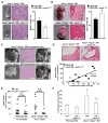
HDL rescue the myeloproliferative disorder and accelerated atherosclerosis of Abca1−/− Abcg1−/− BM transplanted mice. (A) Representative spleen, (B) heart, (C) liver, Peyer’s patches, and hematoxylin and eosin (H&E) staining of paraffin-embedded sections from high cholesterol–fed Ldlr+/ and Ldlr+/− apoA-1 transgenic recipient mice transplanted with Abca1−/− Abcg1−/− BM. (D) Representative H&E staining of the proximal aortas showing atherosclerotic lesions and correlation between mean lesion areas and leukocyte counts. (E) Lesion areas (individual and mean) were determined by morphometric analysis of H&E-stained sections. BM transplanted mice were fed a high-cholesterol diet for different time periods to approximately match lesion areas for mice not expressing apoA-1 transgene. (F) Leukocyte counts from WT and Abca1−/− Abcg1−/− transplanted recipients. Results are means ± SEM (error bars) of six to nine mice per group. *P < 0.05 apoA-I transgene effect; §P < 0.05 BM transplant effect.

Comment in

Similar articles

Cited by

References

    1. Coller BS. Arterioscler Thromb Vasc Biol. 2005;25:658. - PubMed
    1. Gordon DJ, Rifkind BM. N Engl J Med. 1989;321:1311. - PubMed
    1. Barter PJ, Puranik R, Rye KA. Curr Cardiol Rep. 2007;9:493. - PubMed
    1. Tall AR, Yvan-Charvet L, Terasaka N, Pagler T, Wang N. Cell Metab. 2008;7:365. - PubMed
    1. Duffy D, Rader DJ. Nat Rev Cardiol. 2009;6:455. - PubMed

Publication types

MeSH terms

Substances