

Cystatin C increases in cardiac injury: a role in extracellular matrix protein modulation
- PMID: 20489058
- PMCID: PMC2920813
- DOI: 10.1093/cvr/cvq138
Cystatin C increases in cardiac injury: a role in extracellular matrix protein modulation
Abstract
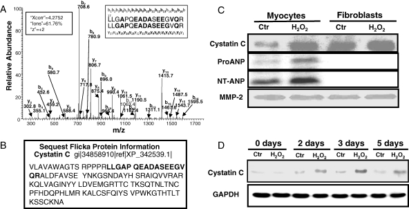
Aims: Numerous lines of evidence suggest a role of oxidative stress in initiation and progression of heart failure. We identify novel pathways of oxidative stress in cardiomyocytes using proteomic technology.
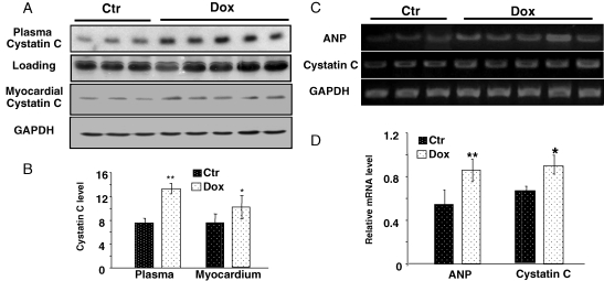
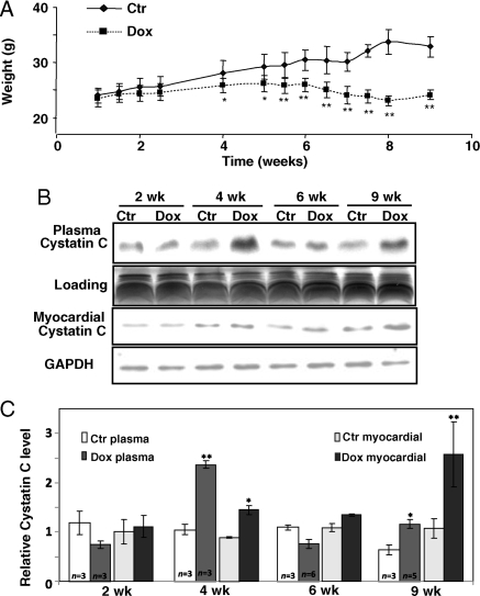
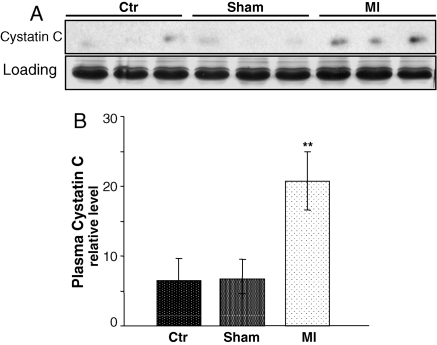
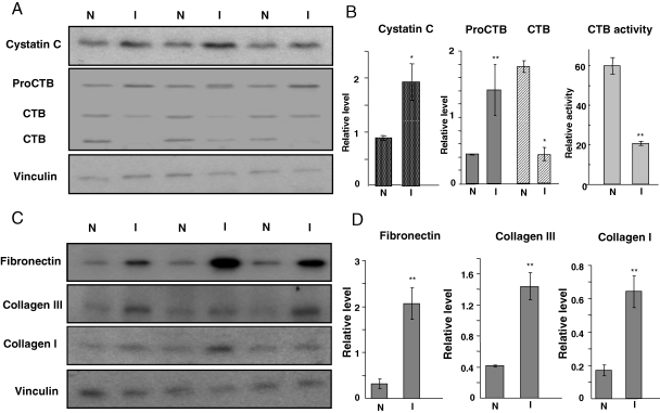
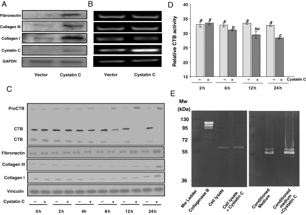
Methods and results: Cardiomyocytes and cardiac fibroblasts isolated from rat hearts were treated with sublethal doses of H(2)O(2) for detection of secreted protein factors in the conditioned media by mass spectrometry-based proteomics. Comparison between the two cell types leads to the finding that H(2)O(2) caused an elevated cystatin C protein in the conditioned medium from cardiomyocytes. When cardiomyopathy was induced in mice by chronic administration of doxorubicin, elevated cystatin C protein was detected in the plasma. Myocardial ischaemia by left anterior descending coronary artery occlusion causes an increase in the level of cystatin C protein in the plasma. In myocardial tissue from the ischaemic area, an increase in cystatin C correlates with the inhibition of cathepsin B activity and accumulation of fibronectin and collagen I/III. Overexpressing cystatin C gene or exposing fibroblasts to cystatin C protein results in an inhibition of cathepsin B and accumulation of fibronectin and collagen I/III.
Conclusion: Oxidants induce elevated cystatin C production from CMCs. Cystatin C plays a role in cardiac extracellular matrix remodelling.
Figures
Comment in
-
Altered degradation of extracellular matrix in myocardial remodelling: the growing role of cathepsins and cystatins.Cardiovasc Res. 2010 Sep 1;87(4):591-2. doi: 10.1093/cvr/cvq208. Epub 2010 Jun 23. Cardiovasc Res. 2010. PMID: 20573730 No abstract available.
Similar articles
-
Altered degradation of extracellular matrix in myocardial remodelling: the growing role of cathepsins and cystatins.Cardiovasc Res. 2010 Sep 1;87(4):591-2. doi: 10.1093/cvr/cvq208. Epub 2010 Jun 23. Cardiovasc Res. 2010. PMID: 20573730 No abstract available.
-
Prenatal Alcohol Exposure Causes Adverse Cardiac Extracellular Matrix Changes and Dysfunction in Neonatal Mice.Cardiovasc Toxicol. 2019 Oct;19(5):389-400. doi: 10.1007/s12012-018-09503-8. Cardiovasc Toxicol. 2019. PMID: 30684169 Free PMC article.
-
Proteome Alterations in Cardiac Fibroblasts: Insights from Experimental Myocardial Infarction and Clinical Ischaemic Cardiomyopathy.Int J Mol Sci. 2025 Apr 18;26(8):3846. doi: 10.3390/ijms26083846. Int J Mol Sci. 2025. PMID: 40332511 Free PMC article.
-
The Extracellular Matrix in Ischemic and Nonischemic Heart Failure.Circ Res. 2019 Jun 21;125(1):117-146. doi: 10.1161/CIRCRESAHA.119.311148. Epub 2019 Jun 20. Circ Res. 2019. PMID: 31219741 Free PMC article. Review.
-
Extracellular matrix remodeling in animal models of anthracycline-induced cardiomyopathy: a meta-analysis.J Mol Med (Berl). 2021 Sep;99(9):1195-1207. doi: 10.1007/s00109-021-02098-8. Epub 2021 May 29. J Mol Med (Berl). 2021. PMID: 34052857 Free PMC article.
Cited by
-
Cystatin C is a disease-associated protein subject to multiple regulation.Immunol Cell Biol. 2015 May-Jun;93(5):442-51. doi: 10.1038/icb.2014.121. Epub 2015 Feb 3. Immunol Cell Biol. 2015. PMID: 25643616 Free PMC article. Review.
-
Association of epicardial adipose tissue with serum level of cystatin C in type 2 diabetes.PLoS One. 2017 Sep 18;12(9):e0184723. doi: 10.1371/journal.pone.0184723. eCollection 2017. PLoS One. 2017. PMID: 28922364 Free PMC article. Clinical Trial.
-
Redox proteins are constitutively secreted by skeletal muscle.J Physiol Sci. 2014 Nov;64(6):401-9. doi: 10.1007/s12576-014-0334-7. Epub 2014 Sep 10. J Physiol Sci. 2014. PMID: 25205643 Free PMC article.
-
Estimation of kidney function in patients with primary neuromuscular diseases: is serum cystatin C a better marker of kidney function than creatinine?J Nephrol. 2022 Mar;35(2):493-503. doi: 10.1007/s40620-021-01122-x. Epub 2021 Aug 5. J Nephrol. 2022. PMID: 34351595 Free PMC article.
-
Managing chemotherapy-related cardiotoxicity in survivors of childhood cancers.Paediatr Drugs. 2014 Oct;16(5):373-89. doi: 10.1007/s40272-014-0085-1. Paediatr Drugs. 2014. PMID: 25134924 Free PMC article. Review.
References
-
- Barth E, Stammler G, Speiser B, Schaper J. Ultrastructural quantitation of mitochondria and myofilaments in cardiac muscle from 10 different animal species including man. J Mol Cell Cardiol. 1992;24:669–681. doi:10.1016/0022-2828(92)93381-S. - DOI - PubMed
-
- Keith M, Geranmayegan A, Sole MJ, Kurian R, Robinson A, Omran AS, et al. Increased oxidative stress in patients with congestive heart failure. J Am Coll Cardiol. 1998;31:1352–1356. doi:10.1016/S0735-1097(98)00101-6. - DOI - PubMed
-
- Singh N, Dhalla AK, Seneviratne C, Singal PK. Oxidative stress and heart failure. Mol Cell Biochem. 1995;147:77–81. doi:10.1007/BF00944786. - DOI - PubMed
-
- Sawyer DB, Colucci WS. Mitochondrial oxidative stress in heart failure: ‘oxygen wastage’ revisited. Circ Res. 2000;86:119–120. - PubMed
-
- Sugden PH, Clerk A. Oxidative stress and growth-regulating intracellular signaling pathways in cardiac myocytes. Antioxid Redox Signal. 2006;8:2111–2124. doi:10.1089/ars.2006.8.2111. - DOI - PubMed
Publication types
MeSH terms
Substances
Grants and funding
LinkOut - more resources
Full Text Sources
Medical
