

Review
doi: 10.2214/AJR.10.4312.
Prostate MRI and 3D MR spectroscopy: how we do it
Affiliations
- PMID: 20489079
- PMCID: PMC2895419
- DOI: 10.2214/AJR.10.4312
Item in Clipboard
Review
Prostate MRI and 3D MR spectroscopy: how we do it
AJR Am J Roentgenol.
2010 Jun.
Abstract
Objective: This review is a primer on the technical aspects of performing a high-quality MRI and MR spectroscopic imaging examination of the prostate.
Conclusion: MRI and MR spectroscopic imaging are useful tools in the localization, staging, and functional assessment of prostate cancer. Performing a high-quality MR spectroscopic examination requires understanding of the technical aspects and limitations of spectral acquisition, postprocessing techniques, and spectral evaluation.
Figures
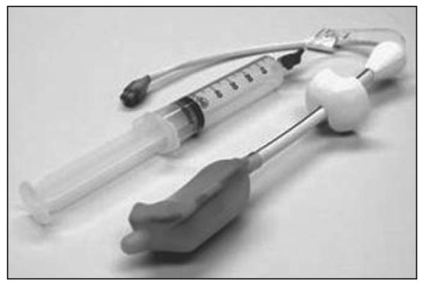
Photograph shows expandable endorectal coil.
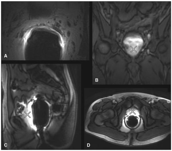
62-year-old man with prostate cancer. A-D, MR images show steps in evaluation of coil position. For optimal coil placement, coil chosen should cover entire prostate (X) (A and B). Signal coverage should be checked from superior to inferior aspect with sagittal fast spin-echo localizer images (C). Anterior to posterior coverage (D) should be checked to make sure coil is not rotated.
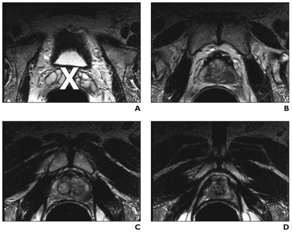
70-year-old man with prostate cancer. A-D, MR images show goals in volume prescription are to cover whole gland, especially peripheral zone, without seminal vesicles; minimize inclusion of air interface; and minimize lipid inclusion. Image A was obtained at level of seminal vesicle (X). Images B–D are regions to be included in volume prescription: B, level of prostate base; C, level of midgland; and D, level of prostate apex.
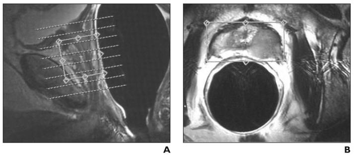
50-year-old man with biopsy-proven prostate cancer of Gleason grade 7 (4 + 3). A and B, Axial (A) and axial oblique (B) MR images show prescription of volume of interest. Volume box should be placed and sized on image.
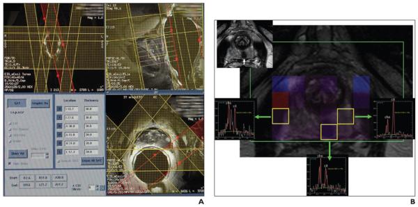
Saturation band placement. A, 60-year-old man with prostate cancer and prostate-specific antigen level of 9.2 ng/dL. Axial T2-weighted localizer MR images show prescription of four very selective suppression radiofrequency bands. Two bands are prescribed on sagittal localizer images. B, 56-year-old man with prostate cancer with serum prostate-specific antigen of 3.3 ng/dL. Pitfall due to low apical periprostatic fat. MR spectroscopic image shows single abnormal voxel with elevated choline to citrate (cho/cit) ratio (0.801) in apical midline peripheral zone. Voxel in left low apical peripheral zone is normal. Voxel on right has elevated cho/cit ratio (0.443) but does not include tumor; elevated cho/cit ratio is secondary to periprostatic fat contamination, which is common pitfall at low apex.
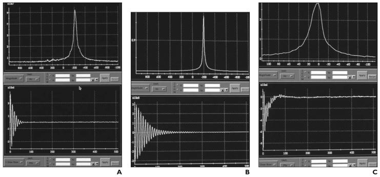
56-year-old man with biopsy-proven prostate cancer. In shimming of spectroscopy window before acquisition, there are typically several display options for evaluating spectra. In this instance, two display windows are used. Fourier transformation takes time domain function (free-induction decay [bottom]) and converts it into frequency domain function (spectrum [top]). A, Example of standard automatic shim spectrum result from entire prostate volume. Top screen shows water peak at–200-Hz off resonance. Shoulder on water peak indicates need for further manual shimming to improve magnetic field homogeneity. It is possible to improve homogeneity through prostate volume by manually adjusting x, y, and z gradient currents. B, Optimized gradient shimming. Improving homogeneity through volume of interest by manual adjustment of x, y, and z gradient currents. With time domain function, it is ideal that decay be as long as possible without evidence of harmonics on display. Goal is to achieve smooth exponential slope of envelope as long in time as possible. C, Recentering water peak exactly on center frequency. Magnitude display is expanded for very precise recentering of water peak.
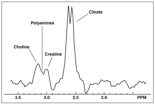
MR spectra of normal human prostate.
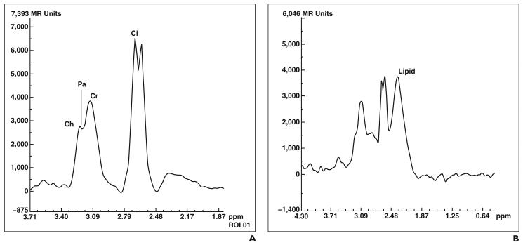
Metabolite peaks in patients with prostate cancer. A, 62-year-old man with prostate cancer of Gleason grade 7 (3 + 4). Ch = choline, Pa = polyamine, Cr = creatine, Ci = citrate. B, 65-year-old man with prostate cancer. Spectra show unwanted lipid signal. Large lipid resonance can obscure prostate metabolite resonance peaks.

Adjustments to spectra. A, Automated baseline correction: sinusoidal curvature of baseline. B, Automated phase correction.
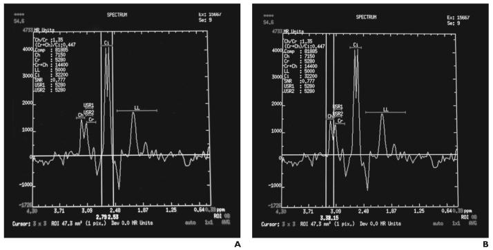
Set ranges for metabolites. A, Citrate (Ci) peak B, Choline (Ch) peak.
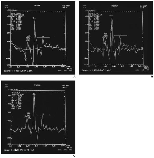
50-year-old man with prostate cancer undergoing MRI for cancer staging. A and B, Spectra prior to baseline (A) and phase correction (B). C, Spectra after baseline and phase correction.
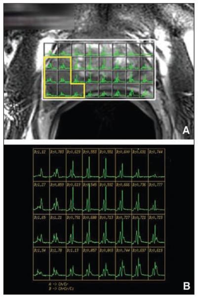
62-year-old man with biopsy-proven prostate cancer with prostate-specific antigen level of 6.8 ng/dL and Gleason grade 6 (3 + 3). A, T2-weighted axial MR image shows 1.7-cm low-signal-intensity mass in right mid gland with prostate capsular bulge. B, Spectroscopy grid from A shows abnormal ratio of choline and creatine to citrate ratio corresponding to low-signal-intensity mass.
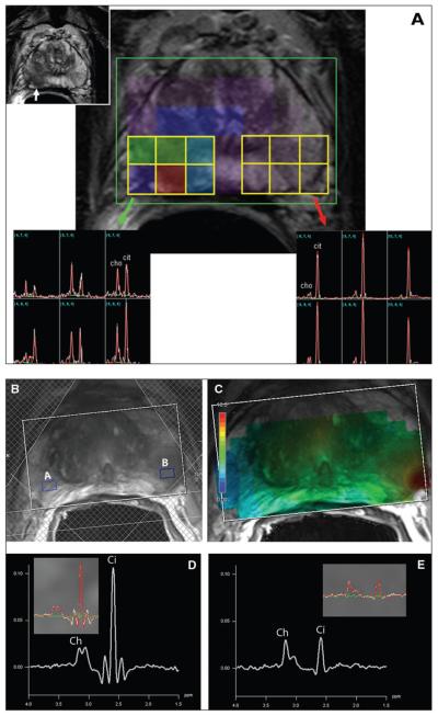
Examples of output seen by radiologist. A, 73-year-old man with prostate cancer with serum prostate-specific antigen level of 45 ng/dL. MR spectroscopic images corresponding to voxels axial T2-weighted MR image show increased choline (cho) to citrate (cit) ratio (green arrow) in right mid peripheral zone tumor (white arrow). Red arrow indicates normal cho/cit ratio in left mid peripheral zone. B–E, 58-year-old man with prostate cancer. Axial 3-T T2-weighted MR images (B and C) show tumor in left peripheral zone. Spectra (D and E) corresponding to partitions (A and B) of MRI image show healthy peripheral zone tissue and tumor. Insets in spectra are automated fits to spectra that quantify signals from choline, creatine, and citrate. These signals can be combined in map of ratio of choline and creatine to citrate.
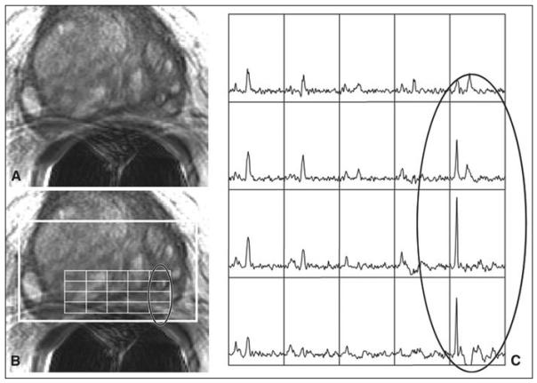
56-year-old man with biopsy-proven prostate cancer. A and B, T2-weighted axial MR images at level of base of prostate (A) and corresponding image (B) with overlaid point-resolved spectroscopy volume (bold white outline) and spectral grid. Oval outline indicates seminal vesicles bleeding into left lateral aspect. C, Spectra (oval) corresponding to oval in B show very high resonance resembling choline peak due to very high glycerophosphocholine level in seminal fluid. This finding is often misinterpreted as prostate cancer.
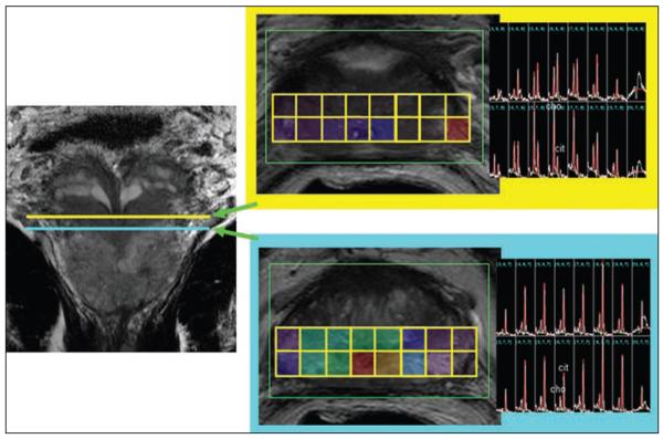
57-year-old man with prostate cancer with serum prostate-specific antigen level of 4.5 ng/dL. Pitfall due to seminal vesicle contamination. MR spectroscopic image shows increased choline-to-citrate ratios in voxels at base due to seminal vesicle contamination (yellow). MR spectroscopic image in more inferior location (turquoise) shows normal choline-to-citrate ratios.
64-year-old man with prostate cancer with prostate-specific antigen level of 7.2 ng/dL and Gleason grade of 6 (3 + 3). A, Axial T1-weighted MR image shows extensive postbiopsy hemorrhage (high signal intensity) on left lobe of prostate 3 weeks after biopsy. B, Axial T2-weighted image corresponding to A shows low signal intensity in same region. C, Spectral array shows loss of spectral signal in region of postbiopsy hemorrhage. Patients have had both metabolic atrophy and changes in metabolite levels in regions of extensive hemorrhage soon after biopsy, confounding ability to metabolically detect prostate cancer. Ch = choline, Cr = creatine, Pa = polyamines.
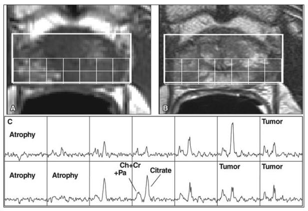
61-year-old man with elevated prostate-specific antigen level of 4.0 ng/dL. A and B, Axial T2-weighted image (A) at mid gland level of prostate and corresponding image with overlaid spectral grid (B) show region of decreased signal intensity (arrows, A) in left lobe consistent with prostate cancer. C, Histopathologic image shows extensive acute inflammation in left lobe of prostate. Biopsy findings were negative for cancer. D, Spectral array corresponding to A and B shows reduction in overall spectral signal but elevated choline to citrate ratio in region of T2 abnormality, also suggesting prostate cancer.
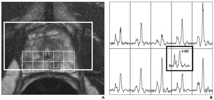
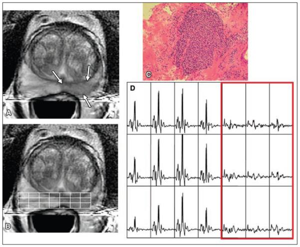
54-year-old man with prostate cancer. A, Axial T2-weighted MR image at level of apex of prostate with overlaid point-resolved spectroscopy box (bold white line) and spectral grid. B, Spectral array corresponding to A shows small region of low signal intensity in left apex corresponds to region of biopsy-proven prostate cancer. Region of low T2 signal intensity is split between two voxels (> 3 SD) in original spectral array. Because volume MRI and MR spectroscopic imaging data are collected, spectroscopic voxels can be shifted in postprocessing to optimally encompass region of abnormality on MR image.
References
-
- Heijmink SW, Futterer JJ, Hambrock T, et al. Prostate cancer: body-array versus endorectal coil MR imaging at 3 T—comparison of image quality, localization, and staging performance. Radiology. 2007;244:184–195. - PubMed
-
- Qayyum A, Coakley FV, Lu Y, et al. Organ-confined prostate cancer: effect of prior transrectal biopsy on endorectal MRI and MR spectroscopic imaging. AJR. 2004;183:1079–1083. - PubMed
-
- Claus FG, Hricak H, Hattery RR. Pretreatment evaluation of prostate cancer: role of MR imaging and 1H MR spectroscopy. RadioGraphics. 2004;24(suppl 1):S167–S180. - PubMed
-
- Coakley FV, Kurhanewicz J, Lu Y, et al. Prostate cancer tumor volume: measurement with endorectal MR and MR spectroscopic imaging. Radiology. 2002;223:91–97. - PubMed
Publication types
MeSH terms
Substances
Grants and funding
LinkOut - more resources
Full Text Sources
Medical
