. 2010 Jul;177(1):70-83.
doi: 10.2353/ajpath.2010.091164. Epub 2010 May 20.

Early growth response-1 induces and enhances vascular endothelial growth factor-A expression in lung cancer cells

Affiliations

Early growth response-1 induces and enhances vascular endothelial growth factor-A expression in lung cancer cells

Hiroaki Shimoyamada et al. Am J Pathol. 2010 Jul.

Abstract

Vascular endothelial growth factor-A (VEGF-A) is crucial for angiogenesis, vascular permeability, and metastasis during tumor development. We demonstrate here that early growth response-1 (EGR-1), which is induced by the extracellular signal-regulated kinase (ERK) pathway activation, activates VEGF-A in lung cancer cells. Increased EGR-1 expression was found in adenocarcinoma cells carrying mutant K-RAS or EGFR genes. Hypoxic culture, siRNA experiment, luciferase assays, chromatin immunoprecipitation, electrophoretic mobility shift assays, and quantitative RT-PCR using EGR-1-inducible lung cancer cells demonstrated that EGR-1 binds to the proximal region of the VEGF-A promoter, activates VEGF-A expression, and enhances hypoxia inducible factor 1alpha (HIF-1alpha)-mediated VEGF-A expression. The EGR-1 modulator, NAB-2, was rapidly induced by increased levels of EGR-1. Pathology samples of human lung adenocarcinomas revealed correlations between EGR-1/HIF-1alpha and VEGF-A expressions and relative elevation of EGR-1 and VEGF-A expression in mutant K-RAS- or EGFR-carrying adenocarcinomas. Both EGR-1 and VEGF-A expression increased as tumors dedifferentiated, whereas HIF-1alpha expression did not. Although weak correlation was found between EGR-1 and NAB-2 expressions on the whole, NAB-2 expression decreased as tumors dedifferentiated, and inhibition of DNA methyltransferase/histone deacetylase increased NAB-2 expression in lung cancer cells despite no epigenetic alteration in the NAB-2 promoter. These findings suggest that EGR-1 plays important roles on VEGF-A expression in lung cancer cells, and epigenetic silencing of transactivator(s) associated with NAB-2 expression might also contribute to upregulate VEGF-A expression.


Figures

Figure 1
Figure 1
Analysis of EGR-1, HIF-1α, VEGF-A, NAB-2, and NAB-1 expression in cultivated lung cancer cells. A: Western blot analysis of whole-cell lysates from adenocarcinoma cell lines containing wild-type K-RAS and EGFR (TKB6 and TKB14), mutant K-RAS (A549, H358), or mutant EGFR (PC9 and HCC827) and from control and EGR-1-transfected TKB5 cells (TKB5pRevEmpty and TKB5pRevEGR-1) and their parental cells (TKB5). Lysates were separated by SDS-PAGE and immunoblotted using antibodies against the proteins indicated. β-Actin was analyzed as an internal control. Lung cancer cells without mutation of K-RAS/EGFR expressed minimal amounts of EGR-1, NAB-2, and VEGF-A, whereas these proteins were readily detectable in mutant K-RAS–carrying cells (A549 and H358) and mutant EGFR-carrying cells (PC9 and HCC827). EGR-1 was abundantly induced by doxycycline treatment for 24 hours in TKB5pRevEGR-1 cells, and NAB-2 and VEGF-A were also promptly induced by EGR-1 induction. NAB-1 was constitutively expressed, but HIF-1α was not detectable in all cell lines. B: Quantitative RT-PCR (qRT-PCR) analysis in TKB5pRevEmpty cells (open bars) and TKB5pRevEGR-1 cells (closed bars) after doxycycline treatment for the indicated times. HIF-1α expression weakly increased after 48 hours, irrespective of EGR-1 induction. However, EGR-1 induction by doxycycline treatment (1 μg/ml final concentration) up-regulated VEGF-A expression, and the expression levels of TKB5pRevEGR-1 cells (72 hours and 96 hours) were further increasing and much higher than those of TKB5pRevEmpty cells. NAB-2 was promptly induced by EGR-1, whereas NAB-1 expression was not altered. C: Association of VEGF-A expression with EGR-1 and HIF-1α. TKB5pRevEGR-1 cells were cultivated in the hypoxic condition (0.5% O2 and 5% CO2) for eight hours. Doxycycline (1 μg/ml final concentration) was added 24 hours before harvest. Lysates were separated by SDS-PAGE and immunoblotted using antibodies against the proteins indicated. β-Actin served as an internal control. Relative expression level of VEGF-A was calculated as follows: (intensity of VEGF-A/intensity of β-actin)/[intensity of VEGF-A (no EGR-1 induction and no hypoxic culture)/intensity of β-actin (no EGR-1 induction and no hypoxic culture)]. VEGF-A was not only induced by EGR-1 and HIF-1α but also enhanced by coexpression of EGR-1 and HIF-1α. D: Alteration of VEGF-A expression by forced repression of EGR-1 in H358 cells. The EGR-1–specific or scrambled control siRNA was treated on H358 cells for 24 hours. β-Actin served as an internal control. Relative expression levels of EGR-1 and VEGF-A were calculated as follows: (intensity of EGR-1 or VEGF-A/intensity of β-actin)/[intensity of EGR-1 or VEGF-A (scrambled siRNA)/intensity of β-actin (scrambled siRNA)]. The VEGF-A expression decreased by EGR-1 repression.
Figure 2
Figure 2
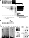
EGR-1 binds to the VEGF-A promoter and activates gene expression. A: VEGF-A promoter assays. Top: TKB5pRevEmpty or TKB5pRevEGR-1 cells were transiently cotransfected with full-length VEGF-A promoter-luciferase reporter construct and control vector for 24 hours with or without doxycycline treatment, and the cells were harvested, lysed, and subjected to luciferase assays. Data reflect means + SD from three experiments performed in triplicate. The mean luciferase activity of full-length VEGF-A promoter in TKB5-pRevEmpty without doxycycline treatment is set to 100%. Bottom: TKB5pRevEGR-1 cells were transiently transfected with wild-type VEGF-A promoter-luciferase reporter construct or the indicated VEGF-A promoter deletion or mutation construct for 24 hours in the presence of doxycycline, and the cells were harvested, lysed, and subjected to luciferase assays. Data reflect the means + SD from three experiments performed in triplicate. The mean luciferase activity of full-length VEGF-A promoter in TKB5-pRevEGR-1 is set to 100%. B: ChIP-based PCR analysis using doxycycline-treated or untreated TKB5pRevEGR-1 cells. DNA fragments conjugated with nuclear proteins were immunoprecipitated with rabbit anti–EGR-1, nonimmunized rabbit IgG, or rabbit anti–HIF-1α antibody. Immunoprecipitated DNA was PCR-amplified using primers for a 377-bp fragment containing the putative EGR-1-binding sites (Table 1) and then separated by agarose gel electrophoresis. Input indicates input DNA; water, no DNA. Weak signal was amplified from the EGR-1 immunoprecipitates even in the condition of no EGR-1 induction (top). This result was consistent with the fact that TKB5pRevEGR-1 constitutively expressed low level of EGR-1–like parental TKB5 cells (Figure 1A). The specific signal was abundantly amplified by EGR-1 induction (bottom). C: EMSA analysis using recombinant EGR-1 (left) and nuclear lysates from TKB5pRevEGR-1 with or without EGR-1 induction (right). Left: Recombinant EGR-1 was incubated with the indicated digoxigenin-labeled EGR-1-binding site probe with or without unlabeled competitor probe (100-fold molar excess) for the consensus EGR-1–binding motif 5′-GCGGGGGCG-3′. EGR-1 bound with highest affinity to sites 1 and 2-4 (arrow). Left, insets: Recombinant EGR-1 was incubated with the indicated digoxigenin-labeled EGR-1–binding site 1 (top) or site 2-4 (bottom) probe with the indicated antibody. Right: Nuclear lysates extracted from EGR-1–induced or uninduced TKB5pRevEGR-1 cells was incubated with the indicated digoxigenin-labeled EGR-1–binding site 1 (top) or site 2-4 (bottom) probe with the indicated antibody. Abundant EGR-1–specific complex was formed by EGR-1 induction and the signal supershifted by anti–EGR-1 antibody. Arrow, EGR-1–specific signal; asterisk, Supershifted EGR-1–specific signal.
Figure 3
Figure 3
Immunohistochemistry of VEGF-A, EGR-1, and HIF-1α expression in surgically resected lung adenocarcinomas. Representative images are shown. A: In a well-differentiated adenocarcinoma, VEGF-A expression is diffuse in the cytoplasm, and EGR-1 is highly expressed in nuclei; HIF-1α is not expressed. B: In a poorly-differentiated adenocarcinoma, VEGF-A expression is diffuse in the cytoplasm, and EGR-1 and HIF-1α are both primarily expressed in nuclei. Scale bars = 100 μm. C: Relationships between EGR-1 and VEGF-A, between HIF-1α and VEGF-A, between EGR-1 and NAB-2, and between EGR-1 and HIF-1α (Pearson’s correlation coefficient test). The expression score (ES) of EGR-1 was correlated with that of VEGF-A and NAB-2, and the ES of HIF-1α was also correlated with that of VEGF-A. However, there was no correlation between EGR-1 and HIF-1α.
Figure 4
Figure 4
NAB-2, EGR-1, and VEGF-A promoter CpG islands are not hypermethylated in lung cancer cells. A: Localization of CpG islands and regions subjected to bisulfite-modified DNA sequencing (BS). Nucleotide positions are relative to the transcription start site. B and C: Summary of BS analysis of NAB-2, EGR-1, and VEGF-A promoter methylation using bisulfite-modified DNA extracted from cultivated lung cancer cells (B) and in vivo adenocarcinoma cells (C). Nucleotide positions are relative to the transcription start site. Closed circle indicates methylated CpG site; open circle, unmethylated CpG site. No severe methylation was noted in NAB-2, EGR-1, or VEGF-A promoters of all cell lines and in vivo adenocarcinoma cells examined. Parenthesis indicates the expression score of each case. W1–W2, M1–M2, and P1–P6 indicated well-differentiated (W), moderately differentiated (M), and poorly differentiated (P) adenocarcinoma samples, respectively. D: Alteration of NAB-2 expression via 5-aza-2′-deoxycytidine treatment in cultivated lung cancer cells. Cells were treated with 10 nmol/ml of 5-aza-2′-deoxycytidine (AZA) for 72 hours or with 300 ng/ml of trichostatin A (TSA) for 24 hours. In addition, cells were also treated with AZA for 48 hours and then with a combination of AZA and TSA for an additional 24 hours. Apparent increase of NAB-2 expression was found in TKB6, TKB14, and H358 cells through the AZA and/or TSA treatment.

References

    1. Ferrara N, Davis-Smith T. The biology of vascular endothelial growth factor. Endocr Rev. 1997;18:4–5. - PubMed
    1. Neufeld G, Cohen T, Gengrinovitch S, Poltorak Z. Vascular endothelial growth factor (VEGF) and its receptors. FASEB J. 1999;13:9–22. - PubMed
    1. Gale NW, Yancopoulos GD. Growth factors acting via endothelial cell-specific receptor tyrosine kinases: vEGFs, angiopoietins, and ephrins in vascular development. Genes Dev. 1999;13:1055–1066. - PubMed
    1. Ferrara N, Gerber HP, LeCouter J. The biology of VEGF and its receptors. Nat Med. 2003;9:669–676. - PubMed
    1. Fontanini G, Vignati S, Boldrini L, Chine S, Silvestri V, Lucchi M, Mussi A, Angeletti CA, Bevilacqua G. Vascular endothelial growth factor is associated with neovascularization and influences progression of non-small cell lung carcinoma. Clin Cancer Res. 1997;3:861–865. - PubMed

Publication types

MeSH terms