. 2010 Jul 23;285(30):23198-207.
doi: 10.1074/jbc.M110.123844. Epub 2010 May 19.

Sequence conversion by single strand oligonucleotide donors via non-homologous end joining in mammalian cells

Affiliations

Sequence conversion by single strand oligonucleotide donors via non-homologous end joining in mammalian cells

Jia Liu et al. J Biol Chem. .

Abstract

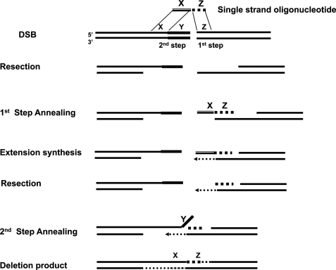
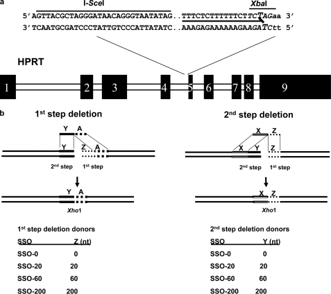
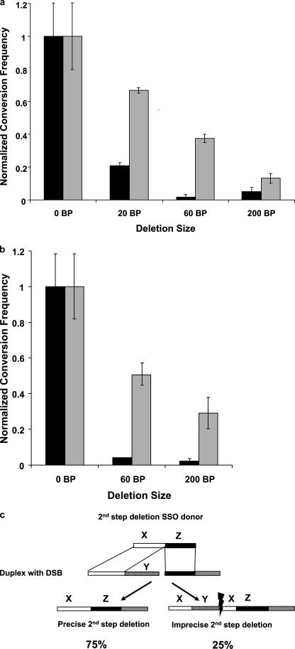
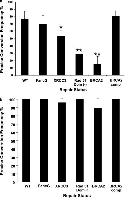
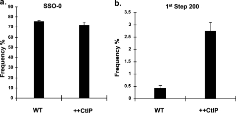
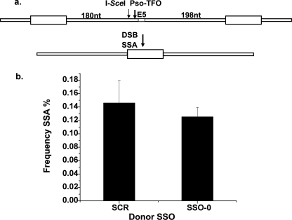
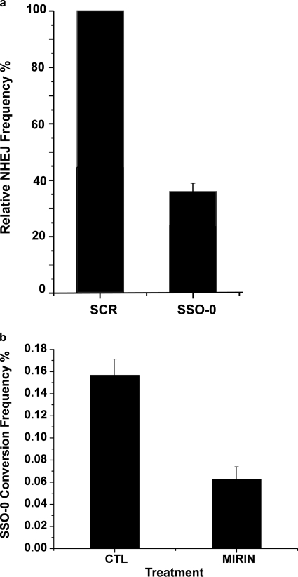
Double strand breaks (DSBs) can be repaired by homology independent nonhomologous end joining (NHEJ) pathways involving proteins such as Ku70/80, DNAPKcs, Xrcc4/Ligase 4, and the Mre11/Rad50/Nbs1 (MRN) complex. DSBs can also be repaired by homology-dependent pathways (HDR), in which the MRN and CtIP nucleases produce single strand ends that engage homologous sequences either by strand invasion or strand annealing. The entry of ends into HDR pathways underlies protocols for genomic manipulation that combine site-specific DSBs with appropriate informational donors. Most strategies utilize long duplex donors that participate by strand invasion. Work in yeast indicates that single strand oligonucleotide (SSO) donors are also active, over considerable distance, via a single strand annealing pathway. We examined the activity of SSO donors in mammalian cells at DSBs induced either by a restriction nuclease or by a targeted interstrand cross-link. SSO donors were effective immediately adjacent to the break, but activity declined sharply beyond approximately 100 nucleotides. Overexpression of the resection nuclease CtIP increased the frequency of SSO-mediated sequence modulation distal to the break site, but had no effect on the activity of an SSO donor adjacent to the break. Genetic and in vivo competition experiments showed that sequence conversion by SSOs in the immediate vicinity of the break was not by strand invasion or strand annealing pathways. Instead these donors competed for ends that would have otherwise entered NHEJ pathways.


Figures

FIGURE 1.
FIGURE 1.
The two-step annealing model for sequence modulation by an SSO donor at a double strand break, based on the schematic of Storici et al. (56). In this illustration the SSO contains homology elements X and Z that are contiguous in the oligonucleotide, but separated by Y in the genome. After introduction of a double strand break at the target site the ends are resected to reveal extended 3′ terminal single strands. The 1st step side (Z) of the SSO donor anneals with the complementary single strand region, and the remainder of the oligonucleotide serves as a template for extension synthesis to generate a new double-stranded end. Additional resection (or oligonucleotide displacement) would reveal an extended 3′ terminal single strand with homology to region X on the other (2nd step) side of the break. Hybridization of the complementary X regions in the 2nd annealing step forms an intermediate that is resolved by elimination of the Y region sequence, fill in, and ligation to yield a precise deletion at the rejoined break.
FIGURE 2.
FIGURE 2.
The 1st and 2nd step SSO deletion donors. a, the pso-TFO and I-SceI target sites in the Chinese hamster Hprt gene in intron 4 adjacent to exon 5. The triplex target site is denoted by a solid line and the cross-linked thymidines are in enlarged font. The first 2 nt of exon 5 are in lowercase. An XbaI site is located immediately adjacent to exon 5. b, schematic of the two kinds of SSO deletion donors. 1st step deletion donors are designed to introduce precise deletions in the region involved in the first annealing step. Conversely 2nd step deletion donors introduce deletions in the region engaged in the 2nd annealing step. The size of the deletion is denoted by the identification of the donor, SSO-60 will introduce a 60-nt deletion, etc. The donors also introduce an XhoI site and eliminate the XbaI site as shown a. In the SSO-0 donor the homology elements immediately adjacent to either side of the break are contiguous to one another, and the conversion product has no deletion.
FIGURE 3.
FIGURE 3.
Activity of 1st and 2nd step deletion donors at targeted DSBs in repair proficient cells. a, frequency (and standard error) of deletion size for 1st step (solid) and 2nd step (gray) deletion SSO donors at a DSB provoked by a targeted cross-link. b, activity of 1st and 2nd step deletion SSO donors at a DSB introduced by I-SceI. c, clones with imprecise products containing partial duplications and deletions of the X and Y sequence elements, as well as clones with precise deletions of the Y region, were recovered.
FIGURE 4.
FIGURE 4.
Activity of SSO donors in cells with deficiencies in FA and HDR genes. a, cells with deficiencies as indicated were transfected with the pso-TFO and the 200-nt 2nd step deletion donor SSO. Hprt-deficient colonies were recovered and the frequency (and standard error) of colonies with precise 200-nt deletions was determined. The statistical significance of the difference between an individual cell line and the wild type cells was p < 0.05 (*) and p < 0.001 (**). b, activity of the SSO-0 donor in cells with deficiencies in FA and HDR genes.
FIGURE 5.
FIGURE 5.
Overexpression of CtIP increases the activity of the 200-nt 1st step deletion donor. Cells were co-electroporated with the CtIP expression plasmid, pso-TFO, and either: a, the SSO-0 donor or, b, the 200-nt 1st step deletion donor. After recovery of Hprt-deficient colonies the frequency of the SSO donor (and standard error) directed product was determined.
FIGURE 6.
FIGURE 6.
Competition between the SSO-0 donor and the SSA pathway. a, schematic of the genomic region containing the SSA construct. The cell line AA8-SSA was constructed by pso-TFO targeted knock-in (“Materials and Methods”) such that a 270-nt sequence in intron 4 was placed in intron 5 as a direct repeat. Resolution by SSA of a DSB introduced by I-SceI, or the targeted cross-link (at arrows), results in a precise deletion of one copy of the repeat, and the sequence between the repeats. This removes exon 5 and inactivates the Hprt gene. b, the AA8-SSA cells were transfected with the pso-TFO and either the SSO-0 or a scrambled (SCR) donor oligonucleotide. The frequency of SSA events was determined by analysis of the target region in Hprt-deficient clones. The standard error for each determination is indicated.
FIGURE 7.
FIGURE 7.
Competition between the SSO-0 donor and the NHEJ pathway. a, cells were co-electroporated with the pso-TFO and either the SSO-0 or a scrambled (SCR) oligonucleotide sequence donor. The frequency of NHEJ was determined in the Hprt-deficient clones. b, cells were co-electroporated with the pso-TFO and SSO-0 donors. Following photoactivation, one aliquot of cells was placed in standard medium containing the same amount of Me2SO as used in the MIRIN-treated sample (CTL). Another was incubated in medium supplemented with 60 μm MIRIN for 20 h, after which the cells were returned to standard medium. The frequency (and standard error) of SSO-0 conversion events was determined.

Similar articles

Cited by

References

    1. Lieber M. R., Lu H., Gu J., Schwarz K. (2008) Cell Res. 18, 125–133 - PubMed
    1. Xie A., Kwok A., Scully R. (2009) Nat. Struct. Mol. Biol. 16, 814–818 - PMC - PubMed
    1. Zha S., Boboila C., Alt F. W. (2009) Nat. Struct. Mol. Biol. 16, 798–800 - PubMed
    1. Capp J. P., Boudsocq F., Bertrand P., Laroche-Clary A., Pourquier P., Lopez B. S., Cazaux C., Hoffmann J. S., Canitrot Y. (2006) Nucleic Acids Res. 34, 2998–3007 - PMC - PubMed
    1. Guirouilh-Barbat J., Rass E., Plo I., Bertrand P., Lopez B. S. (2007) Proc. Natl. Acad. Sci. U.S.A. 104, 20902–20907 - PMC - PubMed

Publication types