. 2010 Jul 23;285(30):23318-30.
doi: 10.1074/jbc.M110.122184. Epub 2010 May 19.

Ligand regulation of the quaternary organization of cell surface M3 muscarinic acetylcholine receptors analyzed by fluorescence resonance energy transfer (FRET) imaging and homogeneous time-resolved FRET

Affiliations

Ligand regulation of the quaternary organization of cell surface M3 muscarinic acetylcholine receptors analyzed by fluorescence resonance energy transfer (FRET) imaging and homogeneous time-resolved FRET

Elisa Alvarez-Curto et al. J Biol Chem. .

Abstract

Flp-In(TM) T-REx(TM) 293 cells expressing a wild type human M(3) muscarinic acetylcholine receptor construct constitutively and able to express a receptor activated solely by synthetic ligand (RASSL) form of this receptor on demand maintained response to the muscarinic agonist carbachol but developed response to clozapine N-oxide only upon induction of the RASSL. The two constructs co-localized at the plasma membrane and generated strong ratiometric fluorescence resonance energy transfer (FRET) signals consistent with direct physical interactions. Increasing levels of induction of the FRET donor RASSL did not alter wild type receptor FRET-acceptor levels substantially. However, ratiometric FRET was modulated in a bell-shaped fashion with maximal levels of the donor resulting in decreased FRET. Carbachol, but not the antagonist atropine, significantly reduced the FRET signal. Cell surface homogeneous time-resolved FRET, based on SNAP-tag technology and employing wild type and RASSL forms of the human M(3) receptor expressed stably in Flp-In(TM) TREx(TM) 293 cells, also identified cell surface dimeric/oligomeric complexes. Now, however, signals were enhanced by appropriate selective agonists. At the wild type receptor, large increases in FRET signal to carbachol and acetylcholine were concentration-dependent with EC(50) values consistent with the relative affinities of the two ligands. These studies confirm the capacity of the human M(3) muscarinic acetylcholine receptor to exist as dimeric/oligomeric complexes at the surface of cells and demonstrate that the organization of such complexes can be modified by ligand binding. However, conclusions as to the effect of ligands on such complexes may depend on the approach used.


Figures

FIGURE 1.
FIGURE 1.
Generation and pharmacological characterization of Flp-InTM T-RExTM293 cells able to express forms of the wild type or RASSL M3 muscarinic acetylcholine receptor on demand. FLAG-hM3WT-Citrine (A and C) or myc-hM3RASSL-Cerulean (B and D) were inserted into the Flp-In locus of Flp-InTM T-RExTM 293 cells, and pools of positive cells were selected. No visible expression of either construct was observed in the absence of the antibiotic doxycycline (A and B, −dox and bright field images), but cell surface delivery of both was produced after addition of doxycycline (A and B, + dox). Elevation of [Ca2+]i in response to varying concentrations of either carbachol (Cch) or clozapine N-oxide (CNO) was assessed in both uninduced and doxycycline-treated cells (C and D), means ± S.E., n = 4.
FIGURE 2.
FIGURE 2.
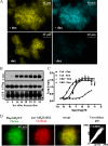
Generation and characterization of Flp-InTM T-RExTM 293 cells able to express inducibly the RASSL M3 muscarinic acetylcholine receptor in the presence of constitutive expression of wild type M3 muscarinic acetylcholine receptor. Cells harboring myc-hM3RASSL-Cerulean at the Flp-In locus as in Fig. 1 (blue) were subsequently further transfected with FLAG-hM3WT-Citrine (yellow), and clones expressing this construct constitutively and stably were isolated. A shows the inducible nature of the RASSL variant (+ dox versusdox). B, membranes from these cells were isolated after treating with doxycycline for varying periods of time, resolved by SDS-PAGE, and immunoblotted with anti-c-Myc (upper panel), anti-FLAG (middle panel), or anti-green fluorescent protein (GFP) (that identifies both cerulean and citrine fluorescent proteins) (lower panel). (Arrowheads show the position of the 105-kDa molecular mass marker.) C, elevation of [Ca2+]i in response to varying concentrations of either carbachol (Cch) or clozapine N-oxide (CNO) was assessed in both RASSL-uninduced (− Dox) and doxycycline-treated cells (+ Dox), means ± S.E., n = 4. D, images of citrine fluorescent proteins (green), cerulean fluorescent proteins (red), and merging of these images (merge) from doxycycline-induced cells were used to construct correlation analyses of color overlap (right-hand picture).
FIGURE 3.
FIGURE 3.
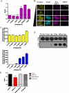
FRET analysis of interactions between variants of the human M3 muscarinic acetylcholine receptor. A and B, co-expression of myc-hM3RASSL-Cerulean (donor) with FLAG-hM3WT-Citrine (acceptor) resulted in raw and corrected (c) FRET signals. Such images were used to calculated ratiometric FRET and how this was modulated by controlling the relative levels of donor to acceptor expression by treatment of cells with varying concentrations of doxycycline. C, levels of acceptor (yellow) and donor (blue) were quantitated at the cell membrane. D, immunological detection of the acceptor (upper panel) and donor (lower panel) at various doxycycline concentrations are displayed (arrowhead indicates the position of the 105-kDa molecular mass marker). E, ratiometric FRET at the cell membrane was measured after treatment with the ligands carbachol, atropine, and clozapine N-oxide subsequent to induction of expression of myc-hM3RASSL-Cerulean with 100 ng·ml−1 of doxycycline (Dox). ***, p < 0.0001. Cch, carbachol; CNO, clozapine N-oxide.
FIGURE 4.
FIGURE 4.
Generation and characterization of Flp-InTM T-RExTM 293 cells able to express SNAP-tagged variants of human M3 muscarinic acetylcholine receptor. A, Flp-InTM T-RExTM 293 cells able to express in an inducible fashion VSV-G-SNAP-hM3WT (left-hand panels) or VSV-G-SNAP-hM3 RASSL (right-hand panels) are shown. Following receptor induction, these were treated with the cell-permeant SNAP substrate SNAP-505 (upper panel and green), an anti-SNAP antibody labeled with Alexa-594 (middle panel and red), or such images were merged (lower panels). Cell nuclei are shown in blue. B, cells induced as in A (top, VSV-G-SNAP-hM3WT; bottom, VSV-G-SNAP-hM3-RASSL) were labeled with the non-cell-permeable reagent Cell-Surface-SNAP-488 and treated with carbachol (1 × 10⁻³ m) or clozapine N-oxide (1 × 10⁻⁴ m) for 0 or 40 min and then imaged. Arrowheads focus attention on receptor populations that became internalized in the presence of ligand. Cch, carbachol; CNO, clozapine N-oxide.
FIGURE 5.
FIGURE 5.
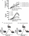
Establishing Tag-LiteTM htrFRET studies to explore receptor dimerization. Cells induced to express differing amounts of VSV-G-SNAP-hM3WT by treatment with varying concentrations of doxycycline (Dox) were incubated with a range of concentrations of the Tb2+-cryptate energy donor SNAP-Lumi4-Tb, and cell surface binding of this ligand was determined by fluorescence intensity at 620 nm (A, top). Using 4 nm SNAP-Lumi4-Tb, varying concentrations of the energy acceptor SNAP-Red were added to cells induced with varying concentrations of doxycycline (A, bottom) to define optimal ratios of energy donor and acceptor to measure receptor homo-interactions. B, concept of the studies is shown for both VSV-G-SNAP-hM3WT and VSV-G-SNAP-hM3RASSL. d2 is a Cisbio trademark for “a second generation htrf acceptor characterized by an organic motif of ∼1,000 Da that is highly FRET compatible with Eu3+ and Tb2+ cryptates.”
FIGURE 6.
FIGURE 6.
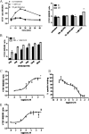
Agonists promote structural re-organization and/or dimerization of the M3 muscarinic acetylcholine receptor. htrFRET studies were conducted in cells induced to express either VSV-G-SNAP-hM3WT or VSV-G-SNAP-hM3RASSL. A, at VSV-G-SNAP-hM3WT carbachol (1 × 10⁻³ m) but not clozapine N-oxide (1 × 10⁻⁴ m) produced a significant increase in basal htrFRET signal, reaching a maximum after 20 min (A). Atropine (1 × 10⁻⁵ m) produced a small but significant decrease in the signal in some but not all studies. B, extent of effect of carbachol was dependent on the level of receptor expression, with substantially greater effects when VSV-G-SNAP-hM3WT expression was lower. C and D, effect of carbachol was concentration-dependent (C) and corresponded to the lower affinity state for carbachol as assessed in competition binding studies employing [3H]QNB (D). E, acetylcholine (Ach) also increased htrFRET signal in a concentration-dependent manner. Cch, carbachol; CNO, clozapine N-oxide.
FIGURE 7.
FIGURE 7.
Clozapine N-oxide promotes structural re-organization and/or enhanced dimerization of the RASSL M3 muscarinic acetylcholine receptor. Clozapine N-oxide produced a concentration-dependent increase of htrFRET signal in cells induced to express VSV-G-SNAP-hM3RASSL after 40 min of treatment, although carbachol (Cch) and atropine (Atro) were without effect.

References

    1. Whorton M. R., Bokoch M. P., Rasmussen S. G., Huang B., Zare R. N., Kobilka B., Sunahara R. K. (2007) Proc. Natl. Acad. Sci. U.S.A. 104, 7682–7687 - PMC - PubMed
    1. Kuszak A. J., Pitchiaya S., Anand J. P., Mosberg H. I., Walter N. G., Sunahara R. K. (2009) J. Biol. Chem. 284, 26732–26741 - PMC - PubMed
    1. Arcemisbéhère L., Sen T., Boudier L., Balestre M. N., Gaibelet G., Detouillon E., Orcel H., Mendre C., Rahmeh R., Granier S., Vivès C., Fieschi F., Damian M., Durroux T., Banères J. L., Mouillac B. (2010) J. Biol. Chem. 285, 6337–6347 - PMC - PubMed
    1. Milligan G. (2008) Br. J. Pharmacol. 153, S216–S229 - PMC - PubMed
    1. Milligan G. (2004) Mol. Pharmacol. 66, 1–7 - PubMed

Publication types

LinkOut - more resources