. 2010 Jul 30;285(31):24099-107.
doi: 10.1074/jbc.M110.128470. Epub 2010 May 19.

Crystal structure of CCM3, a cerebral cavernous malformation protein critical for vascular integrity

Affiliations

Crystal structure of CCM3, a cerebral cavernous malformation protein critical for vascular integrity

Xiaofeng Li et al. J Biol Chem. .

Abstract

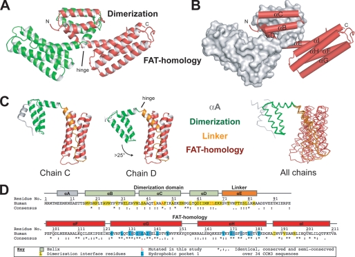
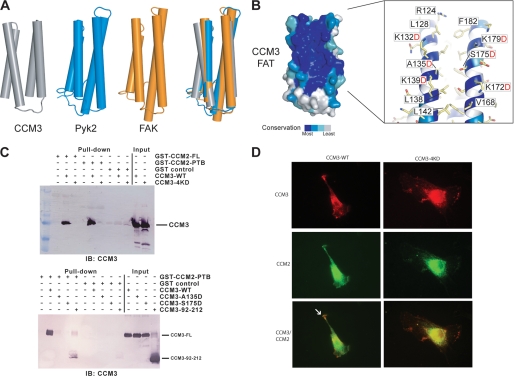
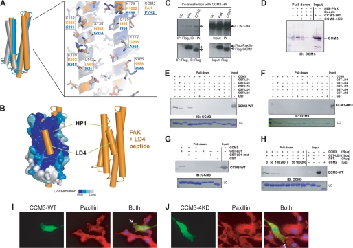
CCM3 mutations are associated with cerebral cavernous malformation (CCM), a disease affecting 0.1-0.5% of the human population. CCM3 (PDCD10, TFAR15) is thought to form a CCM complex with CCM1 and CCM2; however, the molecular basis for these interactions is not known. We have determined the 2.5 A crystal structure of CCM3. This structure shows an all alpha-helical protein containing two domains, an N-terminal dimerization domain with a fold not previously observed, and a C-terminal focal adhesion targeting (FAT)-homology domain. We show that CCM3 binds CCM2 via this FAT-homology domain and that mutation of a highly conserved FAK-like hydrophobic pocket (HP1) abrogates CCM3-CCM2 interaction. This CCM3 FAT-homology domain also interacts with paxillin LD motifs using the same surface, and partial CCM3 co-localization with paxillin in cells is lost on HP1 mutation. Disease-related CCM3 truncations affect the FAT-homology domain suggesting a role for the FAT-homology domain in the etiology of CCM.


Figures

FIGURE 1.
FIGURE 1.
Overall structure of CCM3. A, scheme representation of the CCM3 dimer. The CCM3 dimerization (Dimerization) domain and FAT-homology (FAT-homology) domains are indicated. The location of the hinge between the domains is indicated. B, same orientation as panel A showing one chain as a gray surface and helices αB to αI indicated. C, five solved CCM3 chains hinge around residues Lys-69–Lys-70. Chains C and D of the P212121 crystal form show the largest hinge angle difference and are shown. Backbone trace for all five chains from both crystal forms is superposed on the N-terminal dimerization domain. αA, gray; dimerization domain, green; αE and loop EF, orange; FAT-homology domain, red. D, sequence and secondary structure assignments of human CCM3. Human sequence (Swiss-Prot Q9BUL8) and residue numbers with secondary structure assignment are for helices αA through αI and colored per domain as in panel B. Residues involved in dimerization are colored yellow and the hydrophobic pocket 1 (HP1) in blue. The consensus sequence over 34 CCM3 sequences from human to Drosophila by ClustalW is shown. (*) indicates identical; (:), highly conserved; (.), semi-conserved (see supplemental Fig S4B for complete alignment). Residues mutated in this study are colored red. All structure figures are made with Pymol.
FIGURE 2.
FIGURE 2.
CCM3 dimerization. A, scheme representation of the CCM3 dimer. Exploded views show the anti-parallel hydrophobic stripe for helix αC and the symmetric hydrophobic focal point formed by Ile-66 and Leu-67. B, interacting residues. Surfaces are colored pink for residues that mediate the N-terminal dimerization interface. C, sequence conservation. The surface colored by sequence conservation is based on 34 CCM3 sequences. Darker blue indicates higher conservation. Generated using Consurf (36). D, electrostatic potential representation (+60 kT, blue; −60 kT, red). E, immunoprecipitation. CCM3-HA was co-transfected with Flag-CCM3 variants as indicated into 293T cells. Association of CCM3-HA with full-length CCM3, CCM3 N-terminal region (1–117), and CCM3 FAT domain (92–212) was determined by IP with anti-Flag (M2) followed by Western blot with anti-HA. Flag-CCM3 in the immunoprecipitates was determined by Western blot with anti-Flag. CCM3-HA in the input was also determined. Arrows indicate CCM3. Flag-1–117 is indicated with an (*) and shown in the overexposed panel. VC, vector control. F, size exclusion chromatography. A Superdex 200 analytical grade SEC column was used to analyze CCM3 constructs 1–212 and 92–212. Full-length (1–212) CCM3 elutes as a dimer (predicted mass: ∼25 kDa) and construct 92–212 elutes as a monomer (predicted mass: ∼14 kDa). Standards used to calibrate the column were bovine γ-globulin 158 kDa, chicken ovalbumin 44 kDa, and equine myoglobin 17 kDa. Kav = (VeVo)/(VtVo) where Ve is the elution volume of the protein, Vo is the void volume, and Vt is the total bead volume.
FIGURE 3.
FIGURE 3.
CCM3 FAT-homology domain binds CCM2. A, superposition of CCM3, Pyk2, and FAK. The four helices of Pyk2 FAT domain in blue (PDB ID: 3GM1) (22) and FAK FAT domain in orange (PDB ID: 1OW7) (23) are superposed with the FAT-homology domain of CCM3 (gray). B, surface conservation. The CCM3 FAT-homology domain shows very high conservation on the surfaces of helices αG and αH (see alignment in supplemental Fig. S3). The exploded view labels some of the highly conserved residues and those mutated in this study. C, pull-down. Top: GST fusions of both full-length CCM2 and the predicted CCM2-PTB domain pull-down full-length CCM3. Quadruple mutation of conserved lysines in CCM3, K132D, K139D, K172D, and K179D (CCM3–4KD) renders CCM3 unable to pull-down with CCM2. Bottom: CCM3 HP1 mutations A135D and S175D inhibit pull-down of CCM3 with CCM2-PTB domain. Both full-length CCM3 and CCM3–92-212 pull-down with CCM2-PTB. CCM3 constructs were purified by size exclusion chromatography and eluted as monodisperse peaks. D, immunofluorescence. Either CCM3 FAT-homology domain (lhs) or CCM3 FAT-homology-4KD (rhs) were co-transfected into BAECs with CCM2 and detected by indirect immunofluorescence. CCM2 and CCM3 FAT-homology partially co-localize (white arrow), but introduction of HP1 mutations reduce this co-localization. CCM3 constructs included a C-terminal Flag tag and CCM2, an N-terminal GFP tag.
FIGURE 4.
FIGURE 4.
CCM3 binds the LD-motif region of paxillin. A, left, superposition of CCM3, Pyk2, and FAK as shown in Fig. 3A. Right, Pyk2 colored blue (PDB ID: 3GM1) (22), FAK in orange (PDB ID: 1OW7) (23), and CCM3 (gray). Residues are labeled for comparison. B, superposition of FAK FAT domain bound to paxillin LD4 peptide (PDB ID: 1OW7) (23) with CCM3 FAT-homology domain suggests that CCM3 can bind LD-motifs. C, CCM3 co-immunoprecipitation with CCM2 or paxillin. Full-length CCM3-HA and either full-length Flag-CCM2, full-length Flag-paxillin or vector control (VC) were co-transfected into 293T cells. CCM3 immunoprecipitates both Flag-CCM2 and Flag-paxillin. D, N-terminal 6×His-tagged paxillin-(1–321) pull-down CCM3, but not CCM3–4KD. E–G, CCM3 pull-downs with paxillin. E, GST fusions of paxillin LD-motifs (24, 25) pull-down full-length CCM3. LC, Coomassie-stained loading control for GST-LD motifs. F, quadruple mutation of conserved lysines in CCM3, K132D, K139D, K172D, and K179D (CCM3–4KD) renders CCM3 unable to pull-down with paxillin LD-motifs. G, mutation of paxillin LD1, LL7,8RR, prevents CCM3 pull-down. H, competition. CCM2 and paxillin compete to bind CCM3. Constant GST-LD1 bound to glutathione-Sepharose beads was incubated with constant CCM3 and increasing CCM2 PTB domain. CCM2 PTB domain competes with GST-LD1 for binding to CCM3. I and J, immunofluorescence. Full-length CCM3 (I) or CCM3–4KD (J) were transfected into BAECs. Transfected CCM3 and endogenous paxillin were detected by indirect immunofluorescence microscopy with anti-Flag (rabbit; for CCM3) and anti-paxillin (mouse), followed by FITC-conjugated anti-rabbit and TRITC-conjugated anti-mouse secondary antibodies. CCM3 partially co-localizes with endogenous paxillin at the leading edges of migrating cells. Localization of overexpressed CCM3–4KD to leading edges is reduced (indicated with white arrows).

References

    1. Revencu N., Vikkula M. (2006) J. Med. Genet. 43, 716–721 - PMC - PubMed
    1. Labauge P., Denier C., Bergametti F., Tournier-Lasserve E. (2007) Lancet Neurol. 6, 237–244 - PubMed
    1. Sahoo T., Johnson E. W., Thomas J. W., Kuehl P. M., Jones T. L., Dokken C. G., Touchman J. W., Gallione C. J., Lee-Lin S. Q., Kosofsky B., Kurth J. H., Louis D. N., Mettler G., Morrison L., Gil-Nagel A., Rich S. S., Zabramski J. M., Boguski M. S., Green E. D., Marchuk D. A. (1999) Hum. Mol. Genet. 8, 2325–2333 - PubMed
    1. Günel M., Awad I. A., Anson J., Lifton R. P. (1995) Proc. Natl. Acad. Sci. U.S.A. 92, 6620–6624 - PMC - PubMed
    1. Liquori C. L., Berg M. J., Siegel A. M., Huang E., Zawistowski J. S., Stoffer T., Verlaan D., Balogun F., Hughes L., Leedom T. P., Plummer N. W., Cannella M., Maglione V., Squitieri F., Johnson E. W., Rouleau G. A., Ptacek L., Marchuk D. A. (2003) Am. J. Hum. Genet. 73, 1459–1464 - PMC - PubMed

Publication types