

Ventricle Boundary in CT: Partial Volume Effect and Local Thresholds
- PMID: 20490355
- PMCID: PMC2872763
- DOI: 10.1155/2010/674582
Ventricle Boundary in CT: Partial Volume Effect and Local Thresholds
Abstract
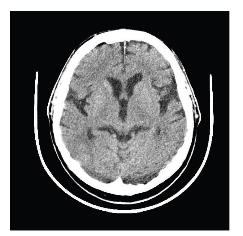
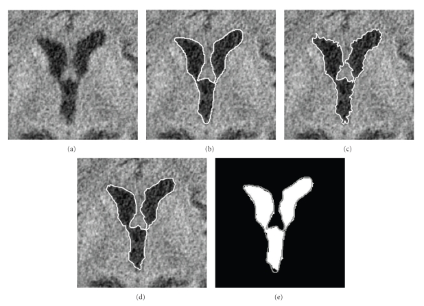
We present a mathematical frame to carry out segmentation of cerebrospinal fluid (CSF) of ventricular region in computed tomography (CT) images in the presence of partial volume effect (PVE). First, the image histogram is fitted using the Gaussian mixture model (GMM). Analyzing the GMM, we find global threshold based on parameters of distributions for CSF, and for the combined white and grey matter (WGM). The parameters of distribution of PVE pixels on the boundary of ventricles are estimated by using a convolution operator. These parameters are used to calculate local thresholds for boundary pixels by the analysis of contribution of the neighbor pixels intensities into a PVE pixel. The method works even in the case of an almost unimodal histogram; it can be useful to analyze the parameters of PVE in the ground truth provided by the expert.
Figures

Similar articles
-
Methods in quantitative image analysis.Histochem Cell Biol. 1996 May;105(5):333-55. doi: 10.1007/BF01463655. Histochem Cell Biol. 1996. PMID: 8781988 Review.
-
An extension Gaussian mixture model for brain MRI segmentation.Annu Int Conf IEEE Eng Med Biol Soc. 2014;2014:4711-4. doi: 10.1109/EMBC.2014.6944676. Annu Int Conf IEEE Eng Med Biol Soc. 2014. PMID: 25571044
-
Efficient Johnson-SB Mixture Model for Segmentation of CT Liver Image.J Healthc Eng. 2022 Apr 14;2022:5654424. doi: 10.1155/2022/5654424. eCollection 2022. J Healthc Eng. 2022. PMID: 35463693 Free PMC article.
-
Positron emission tomography metabolic data corrected for cortical atrophy using magnetic resonance imaging.Alzheimer Dis Assoc Disord. 1996 Fall;10(3):141-70. doi: 10.1097/00002093-199601030-00005. Alzheimer Dis Assoc Disord. 1996. PMID: 8876777
-
Automatic segmentation of cerebrospinal fluid, white and gray matter in unenhanced computed tomography images.Acad Radiol. 2010 Nov;17(11):1350-8. doi: 10.1016/j.acra.2010.06.005. Epub 2010 Jul 15. Acad Radiol. 2010. PMID: 20634108
Cited by
-
Automated delineation of stroke lesions using brain CT images.Neuroimage Clin. 2014 Mar 21;4:540-8. doi: 10.1016/j.nicl.2014.03.009. eCollection 2014. Neuroimage Clin. 2014. PMID: 24818079 Free PMC article.
-
Quantitative estimation of a ratio of intracranial cerebrospinal fluid volume to brain volume based on segmentation of CT images in patients with extra-axial hematoma.Neuroradiol J. 2017 Feb;30(1):10-14. doi: 10.1177/1971400916678227. Epub 2016 Nov 11. Neuroradiol J. 2017. PMID: 27837185 Free PMC article.
-
Validated automatic brain extraction of head CT images.Neuroimage. 2015 Jul 1;114:379-85. doi: 10.1016/j.neuroimage.2015.03.074. Epub 2015 Apr 7. Neuroimage. 2015. PMID: 25862260 Free PMC article.
References
-
- Sezgin M, Sankur B. Survey over image thresholding techniques and quantitative performance evaluation. Journal of Electronic Imaging. 2004;13(1):146–168.
-
- Otsu N. A threshold selection method from gray-level histogram. IEEE Transactions on Systems Man and Cybernetics. 1979;8(1):62–66.
-
- Liao P-S, Chen T-S, Chung P-C. A fast algorithm for multilevel thresholding. Journal of Information Science and Engineering. 2001;17(5):713–727.
-
- Wang H, Dong Y. An improved image segmentation algorithm based on otsu method. In: Zhou L, editor. In: International Symposium on Photoelectronic Detection and Imaging 2007: Related Technologies and Applications, vol. 6625; 2008; pp. 66250I-1–66250I-8. Proceedings of SPIE.
-
- Doyle W. Operations useful for similarity-invariant pattern recognition. Journal of the ACM. 1962;9:259–267.
LinkOut - more resources
Full Text Sources
