

Understanding the formation of maxillary sinus in Japanese human foetuses using cone beam CT
- PMID: 20490493
- PMCID: PMC2945628
- DOI: 10.1007/s00276-010-0678-5
Understanding the formation of maxillary sinus in Japanese human foetuses using cone beam CT
Abstract
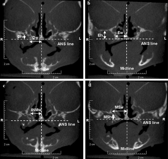
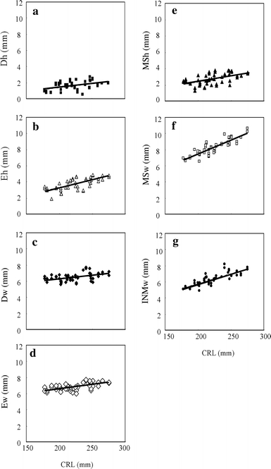
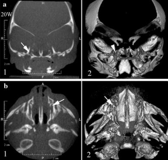
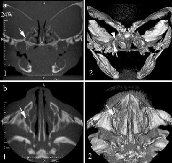
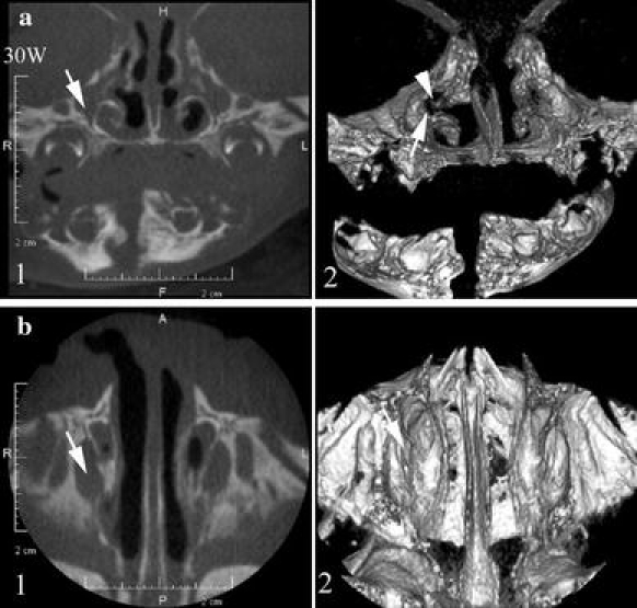
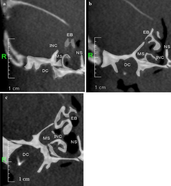
The formation of the maxillary sinus (MS) is tied to the maturation of the craniofacial bones during development. The MS and surrounding bone matrices in Japanese foetal specimens were inspected using cone beam computed tomography relative to the nasal cavity (NC) and the surrounding bones, including the palatine bone, maxillary process, inferior nasal concha and lacrimal bone. The human foetuses analysed were 223.2 ± 25.9 mm in crown-rump length (CRL) and ranged in estimated age from 20 to 30 weeks of gestation. The amount of bone in the maxilla surrounding the MS increased gradually between 20 and 30 weeks of gestation. Various calcified structures that formed the bone matrix were found in the cortical bone of the maxilla, and these calcified structures specifically surrounded the deciduous tooth germs. By 30 weeks of gestation, the uncinate process of the ethmoid bone formed a border with the maxilla. The distance from the midline to the maximum lateral surface border of the MS combined with the width from the midline to the maximum lateral surface border of the inferior nasal concha showed a high positive correlation with CRL in Japanese foetuses. There appears to be a complex correlation between the MS and NC formation during development in the Japanese foetus. Examination of the surrounding bone indicated that MS formation influences maturation of the maxilla and the uncinate process of the ethmoid bone during craniofacial bone development.
Figures
Similar articles
-
Evaluation of the blood and nerve supply patterns in the molar region of the maxillary sinus in Japanese cadavers.Okajimas Folia Anat Jpn. 2010 Nov;87(3):129-33. doi: 10.2535/ofaj.87.129. Okajimas Folia Anat Jpn. 2010. PMID: 21174942
-
Anatomical Changes in a Case with Asymmetrical Bilateral Maxillary Sinus Hypoplasia.Medicina (Kaunas). 2022 Apr 20;58(5):564. doi: 10.3390/medicina58050564. Medicina (Kaunas). 2022. PMID: 35629981 Free PMC article.
-
Distances of root apices to adjacent anatomical structures in the anterior maxilla: an analysis using cone beam computed tomography.Clin Oral Investig. 2019 May;23(5):2253-2263. doi: 10.1007/s00784-018-2650-4. Epub 2018 Oct 4. Clin Oral Investig. 2019. PMID: 30288606
-
Immunocytochemical study of the maxilla and maxillary sinus during human fetal development.Okajimas Folia Anat Jpn. 1998 Oct;75(4):205-16. doi: 10.2535/ofaj1936.75.4_205. Okajimas Folia Anat Jpn. 1998. PMID: 9871404
-
Anatomical structures in the maxillary sinus related to lateral sinus elevation: a cone beam computed tomographic analysis.Clin Oral Implants Res. 2013 Aug;24 Suppl A100:75-81. doi: 10.1111/j.1600-0501.2011.02378.x. Epub 2011 Dec 8. Clin Oral Implants Res. 2013. PMID: 22150785
Cited by
-
Assessment of the Relationship between Maxillary Posterior Teeth and Maxillary Sinus Using Cone-Beam Computed Tomography.Int J Dent. 2022 Jul 5;2022:6254656. doi: 10.1155/2022/6254656. eCollection 2022. Int J Dent. 2022. PMID: 35847346 Free PMC article.
-
Topography of the nasolacrimal duct on the lateral nasal wall in Koreans.Surg Radiol Anat. 2012 Apr;34(3):249-55. doi: 10.1007/s00276-011-0858-y. Epub 2011 Jul 28. Surg Radiol Anat. 2012. PMID: 21796405
-
Evaluation of the lacrimal recess of the maxillary sinus: an anatomical study.Braz J Otorhinolaryngol. 2013 Jan-Feb;79(1):35-8. doi: 10.5935/1808-8694.20130007. Braz J Otorhinolaryngol. 2013. PMID: 23503905 Free PMC article.
-
Accuracy and precision of manual segmentation of the maxillary sinus in MR images-a method study.Br J Radiol. 2018 May;91(1085):20170663. doi: 10.1259/bjr.20170663. Epub 2018 Mar 20. Br J Radiol. 2018. PMID: 29419324 Free PMC article.
-
Quantitative and qualitative bone analysis in the maxillary lateral region.Surg Radiol Anat. 2012 Aug;34(6):551-8. doi: 10.1007/s00276-012-0955-6. Epub 2012 Mar 22. Surg Radiol Anat. 2012. PMID: 22437467
References
MeSH terms
LinkOut - more resources
Full Text Sources
