Review
. 2010 Oct;67(20):3389-405.
doi: 10.1007/s00018-010-0399-2. Epub 2010 May 20.

β-Glucosidases

Affiliations
Review

β-Glucosidases

James R Ketudat Cairns et al. Cell Mol Life Sci. 2010 Oct.

Abstract

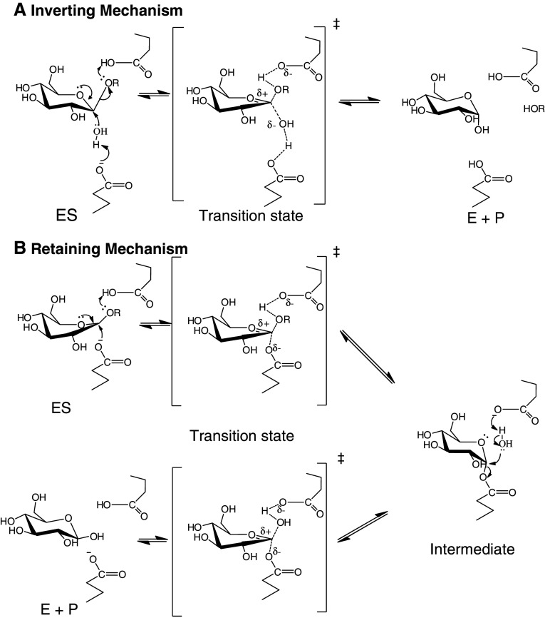
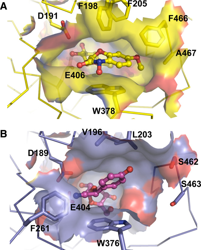
β-Glucosidases (3.2.1.21) are found in all domains of living organisms, where they play essential roles in the removal of nonreducing terminal glucosyl residues from saccharides and glycosides. β-Glucosidases function in glycolipid and exogenous glycoside metabolism in animals, defense, cell wall lignification, cell wall β-glucan turnover, phytohormone activation, and release of aromatic compounds in plants, and biomass conversion in microorganisms. These functions lead to many agricultural and industrial applications. β-Glucosidases have been classified into glycoside hydrolase (GH) families GH1, GH3, GH5, GH9, and GH30, based on their amino acid sequences, while other β-glucosidases remain to be classified. The GH1, GH5, and GH30 β-glucosidases fall in GH Clan A, which consists of proteins with (β/α)(8)-barrel structures. In contrast, the active site of GH3 enzymes comprises two domains, while GH9 enzymes have (α/α)(6) barrel structures. The mechanism by which GH1 enzymes recognize and hydrolyze substrates with different specificities remains an area of intense study.


Figures

Fig. 1
Fig. 1
Structures of example β-glucosidase substrates. The plant cyanogenic glucosides linamarin, dhurrin, prunasin, and its precursor amygdalin. Other defense-related glycosides include 2-O-β-d-glucopyranosyl-4-hydroxy-7-methoxy-1,4-benzoxazin-3-one (DIMBOAGlc) and the flavonoids apigenin 7-O-β-d-glucoside, the isoflavonoids diadzin and genistin, and phloridzin. Coniferin and coumaryl alcohol represent monolignol β-glucosides, while abscissic acid glucosyl ester is a phytohormone glucoconjugate and salicin and indoxyl β-glucoside are other plant glycosides with similarity to phytohormones. Strictosidine is the metabolic precursor to a wide array of monoterpene alkaloids. Cellobiose and laminaribiose represent plant cell-wall-derived oligosaccharides and can be extended with the same linkage to give the corresponding triose, tetraose, etc. In the lower right is an example of a glucosyl ceramide, one of the substrates for human acid β-glucosidase (GBA1) and other mammalian β-glucosidases
Fig. 2
Fig. 2
Structures of β-glucosidases from different GH families. These include β-glucosidases or related enzymes from GH1 (Zea mays ZmGlu1, PDB code 1E1E), GH3 (Hordeum vulgare Exo I β-glucan glucohydrolase, PDB code 1EX1), GH5 (Candida albicans exo-β-(1,3)-glucanase Exg exoglucanase, PDB code 1CZ1), GH30 (Homo sapiens, acid β-glucosidase/glucocerebrosidase GBA1, PDB code 2V3D), and GH9 (Vibrio parahaemolyticus, putative exoglucanase, PDB code 3H7L). The structural cartoons are colored in a spectrum from blue to red from their N- to C-termini, with the catalytic nucleophile and acid–base residues shown in stick for those enzymes in which they are known. The ligands shown are glucose in the GH3 barley ExoI and N-butyl-deoxynojirimycin in the GH30 human GBA1, both of which are shown with carbons in pink. The human GBA2 (bile acid β-glucosidase) shows low levels of sequence similarity to (α/α)6 enzymes, suggesting its catalytic domain may be similar to the GH9 structure. Drawn with Pymol (DeLano)
Fig. 3
Fig. 3
Retaining catalytic mechanisms of inverting and retaining β-glucosidases. a The inverting mechanism that is seen in family GH9 glycoside hydrolases, including β-glucosidases. A single displacement of the aglycone by the water leads to an anomeric carbon with inverted chirality. b The commonly accepted mechanism for hydrolysis with retention of anomeric configuration as seen GH Clan A and family GH3 β-glucosidases. The glucosyl moiety is distorted into an 1S3 skew boat upon binding to the enzyme in preparation to form the 4H3 half chair conformation of the proposed transition state [107, 108]. The first step is glycosylation, in which the catalytic acid donates a proton to the leaving group, while the catalytic nucleophile attacks from the opposite side to form an α-linked intermediate. In the second, deglycosylation step, the catalytic base (the same carboxylate as the catalytic acid) extracts a proton from a water molecule, improving its nucleophilic power to attack at the anomeric carbon and displace the enzyme. Hydrolysis by either mechanism is equivalent in the organism, since mutarotation of the released glucose will lead to a racemic mixture of glucose in solution after a short time
Fig. 4
Fig. 4
Active site configurations of maize β-glucosidase 1 (Glu1, a) and sorghum dhurrinase 1 (Dhr1, b). The active sites of maize Glu1 and sorghum Dhr1 enzymes are shown for the structures of the Glu1 E189D mutant in complex with DIMBOA glucoside (PDB entry 1E56) and Dhr1 E191D mutant in complex with dhurrin (PDB entry 1VO3) [109, 111]. The sidechains of residues noted to interact with the aglycone are shown in stick representation behind the active site surface, which is colored as the underlying residues, which are colored with carbon in yellow for Glu1 and purple for Dhr1, nitrogen in blue, and oxygen in red. The ligands are shown in ball and stick representation with similar coloration. The Phe 261 residue, which narrows the active site in Dhr1, is also shown in front of the catalytic nucleophile Glu 404. Figure created with Pymol

Similar articles

Cited by

References

    1. Henrissat B. A classification of glycosyl hydrolases based on amino acid sequence similarities. Biochem J. 1991;280:309–316. - PMC - PubMed
    1. Henrissat B, Davies G. Structural and sequence-based classification of glycosyl hydrolases. Curr Opin Struct Biol. 1997;7:637–644. doi: 10.1016/S0959-440X(97)80072-3. - DOI - PubMed
    1. Cantarel BL, Coutinho PM, Rancurel C, Bernard T, Lombard V, Henrissat B. The Carbohydrate-Active EnZymes database (CAZy): an expert resource for glycogenomics. Nucleic Acids Res. 2009;37:D233–D238. doi: 10.1093/nar/gkn663. - DOI - PMC - PubMed
    1. Brunner R, Wirtz W, Rose JKC, Darbill AG, Govers F, Scheel D, Nurnberger T. A β-glucosidase/xylosidase from the phytopathogenic oomycete, Phytophthora infestans . Phytochemistry. 2002;59:689–696. doi: 10.1016/S0031-9422(02)00045-6. - DOI - PubMed
    1. Opassiri R, Pomthong B, Akiyama T, Nakphaichit M, Onkoksoong T, Ketudat-Cairns M, Ketudat Cairns JR. A stress-induced rice β-glucosidase represents a new subfamily of glycosyl hydrolase family 5 containing a fascin-like domain. Biochem J. 2007;408:241–249. doi: 10.1042/BJ20070734. - DOI - PMC - PubMed

Publication types

LinkOut - more resources