

Development of a method for effective amplification of human adenovirus 40
- PMID: 20490608
- PMCID: PMC4569093
- DOI: 10.1007/s00705-010-0683-3
Development of a method for effective amplification of human adenovirus 40
Abstract
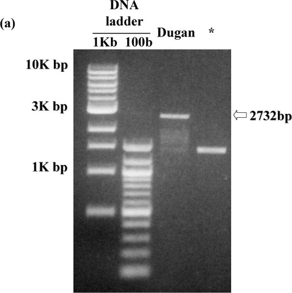
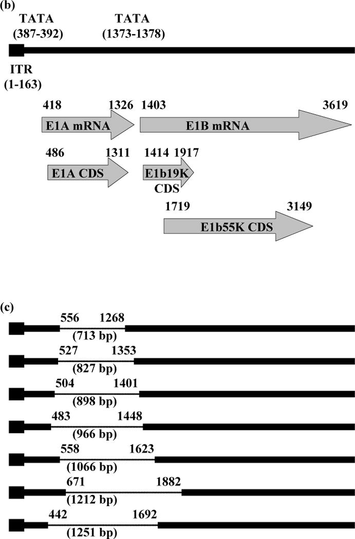
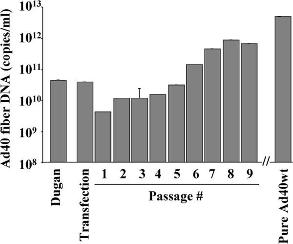
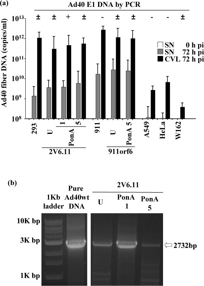
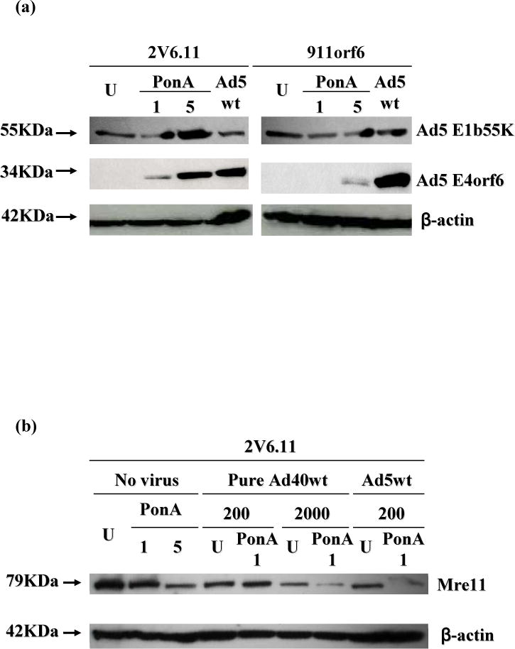
Human adenovirus 40 (Ad40) is an interesting candidate for vector construction because of its tropism for the gastrointestinal tract. Although effective preparation of the vector is necessary for its in vivo application, amplification of Ad40 has been very difficult. Ad40 E1 deletion mutants were detected by PCR in the viral DNA from Ad40 Dugan amplified by Ad5 E1-expressing human embryonic kidney (293) cells and in Ad40 Dugan plaques observed with Ad5 E1-expressing human retinoblastic cells. For the purpose of generating a single wild-type Ad40 clone, the entire Ad40 DNA was cloned into a plasmid by homologous recombination. A pure Ad40 was successfully generated by plasmid transfection and subsequently amplified with Ad5 E4orf6-inducible 293 (2V6.11) cells. 2V6.11 is an apposite cell line for effective Ad40 amplification and for future vector construction because Ad40 genetic integrity was maintained with this Ad5 E1 and E4orf6 trans-complementing cell line.
Figures
Similar articles
-
Molecular characterization of replication-competent variants of adenovirus vectors and genome modifications to prevent their occurrence.J Virol. 1996 Dec;70(12):8459-67. doi: 10.1128/JVI.70.12.8459-8467.1996. J Virol. 1996. PMID: 8970968 Free PMC article.
-
Complementation of enteric adenovirus type 40 for lytic growth in tissue culture by E1B 55K function of adenovirus types 5 and 12.Virology. 1989 Aug;171(2):619-22. doi: 10.1016/0042-6822(89)90634-x. Virology. 1989. PMID: 2527440
-
Designing E1 deleted adenoviral vector by homologous recombination.Iran Biomed J. 2007 Jul;11(3):199-202. Iran Biomed J. 2007. PMID: 18051781
-
Human adenovirus cloning vectors based on infectious bacterial plasmids.Gene. 1986;50(1-3):161-71. doi: 10.1016/0378-1119(86)90321-5. Gene. 1986. PMID: 3472993
-
Development and assessment of human adenovirus type 11 as a gene transfer vector.J Virol. 2005 Apr;79(8):5090-104. doi: 10.1128/JVI.79.8.5090-5104.2005. J Virol. 2005. PMID: 15795294 Free PMC article.
Cited by
-
Species D human adenovirus type 9 exhibits better virus-spread ability for antitumor efficacy among alternative serotypes.PLoS One. 2014 Feb 4;9(2):e87342. doi: 10.1371/journal.pone.0087342. eCollection 2014. PLoS One. 2014. PMID: 24503714 Free PMC article.
-
Intravenous genetic mesothelin vaccine based on human adenovirus 40 inhibits growth and metastasis of pancreatic cancer.Int J Cancer. 2013 Jul;133(1):88-97. doi: 10.1002/ijc.27983. Epub 2012 Dec 21. Int J Cancer. 2013. PMID: 23233329 Free PMC article.
-
Pathogenicity and virulence of human adenovirus F41: Possible links to severe hepatitis in children.Virulence. 2023 Dec;14(1):2242544. doi: 10.1080/21505594.2023.2242544. Virulence. 2023. PMID: 37543996 Free PMC article. Review.
-
A single intraduodenal administration of human adenovirus 40 vaccine effectively prevents anaphylactic shock.Clin Vaccine Immunol. 2013 Oct;20(10):1508-16. doi: 10.1128/CVI.00417-13. Epub 2013 Jul 24. Clin Vaccine Immunol. 2013. PMID: 23885027 Free PMC article.
References
-
- Carter MJ. Enterically infecting viruses: pathogenicity, transmission and significance for food and waterborne infection. J Appl Microbiol. 2005;98:1354–1380. - PubMed
-
- Thorner AR, Vogels R, Kaspers J, Weverling GJ, Holterman L, Lemckert AA, Dilraj A, McNally LM, Jeena PM, Jepsen S, Abbink P, Nanda A, Swanson PE, Bates AT, O’Brien KL, Havenga MJ, Goudsmit J, Barouch DH. Age dependence of adenovirus-specific neutralizing antibody titers in individuals from sub-Saharan Africa. J Clin Microbiol. 2006;44:3781–3783. - PMC - PubMed
Publication types
MeSH terms
Substances
Grants and funding
LinkOut - more resources
Full Text Sources
Other Literature Sources
