. 2010 Nov;106(1-2):3-17.
doi: 10.1007/s11120-010-9555-7. Epub 2010 May 20.

Phylogenomic analysis of the Chlamydomonas genome unmasks proteins potentially involved in photosynthetic function and regulation

Affiliations

Phylogenomic analysis of the Chlamydomonas genome unmasks proteins potentially involved in photosynthetic function and regulation

Arthur R Grossman et al. Photosynth Res. 2010 Nov.

Abstract

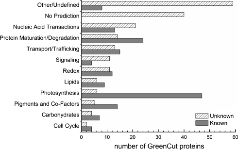
Chlamydomonas reinhardtii, a unicellular green alga, has been exploited as a reference organism for identifying proteins and activities associated with the photosynthetic apparatus and the functioning of chloroplasts. Recently, the full genome sequence of Chlamydomonas was generated and a set of gene models, representing all genes on the genome, was developed. Using these gene models, and gene models developed for the genomes of other organisms, a phylogenomic, comparative analysis was performed to identify proteins encoded on the Chlamydomonas genome which were likely involved in chloroplast functions (or specifically associated with the green algal lineage); this set of proteins has been designated the GreenCut. Further analyses of those GreenCut proteins with uncharacterized functions and the generation of mutant strains aberrant for these proteins are beginning to unmask new layers of functionality/regulation that are integrated into the workings of the photosynthetic apparatus.


Figures

Fig. 1
Fig. 1
Classification of GreenCut proteins. The bar graph presents the functional classification of GreenCut proteins (on y axis) and the number of proteins placed into each class (x axis). The solid bars represent proteins with known functions while the hatched bars represent proteins of unknown functions. The proteins of unknown function have been placed in generalized functional categories based on domains or motifs within the proteins
Fig. 2
Fig. 2
Co-expression of genes of the ATP synthase operon with CGLD22 (sll1321) in Synechocystis sp. PCC 6803. a The microarray data used to generate the expression curves were obtained from the Gene Expression Omnibus (http://www.ncbi.nlm.nih.gov/geo/). The atp1 gene is the putative ortholog of CGLD22; the curve showing the expression profile of atp1 is in red. The curves showing the expression profiles of slr1413 and sll0216, genes that are not part of the ATP synthase operon and were used as a control for these analyses, are a dotted and broken line, respectively. The curves showing expression profiles of all other genes of the ATP synthase operon are in gray. Microarray values were background-corrected, normalized against the median of the ratio of each sample against the reference, and log-transformed. The plotted data include microarray replicates of 38 biological experiments. b The arrangement of genes of the ATP synthase operon. The genes are depicted as arrows, with the orientation indicated by the direction of the arrow. The location of the genes on the chromosome relative to the origin is indicated. This information was obtained from CyanoBase (http://genome.kazusa.or.jp/cyanobase/) (Nakao et al. 2010). The genes of the operon are atp1 (sll1321), atpI (sll1322), atpH (ssl2615), atpG (sll1323), atpF (sll1324), atpD (sll1325), atpA (sll1326), and atpC (sll1327). slr1413 is upstream, and slr1411 and sll0216 are downstream of the ATP synthase operon, respectively, and neither is co-expressed with atp1. All of the genes of the ATP synthase operon are depicted as light gray-filled arrows, except for atp1; this arrow is red-filled. Arrows representing genes outside the operon, slr1411, slr1413, and sll0216, are unfilled and dark gray-filled
Fig. 3
Fig. 3
Analyses of mutants defective for genes encoding GreenCut proteins. Step 1: Mutants are grown at varying light intensities on medium containing acetate or in minimal medium supplemented with CO2. In this example, a strain with a mutation in the CGL28 gene (red box, step 1) grew slower than wild-type cells (blue box) on acetate-containing medium, and did not grow at all on minimal medium supplemented with CO2. Step 2: Fv/Fm values, shown as a false color image, are determined for colonies grown on solid medium containing acetate. In this case, the cgl28 mutant (red box) was determined to have a markedly reduced Fv/Fm relative to wild-type cells (blue box). Step 3: The mutants are further analyzed after growth in the dark in liquid medium containing acetate for photochemical and non-photochemical quenching using fluorescence assays. This strain (blue curve) has no variable fluorescence (which can be observed in the pink curve of wild-type [WT] cells). When the horizontal bar at the top of the image is unfilled (white, outlined in black), the sample is being exposed to actinic light, while the black-filled region of the bar indicates that the sample is in the dark. All downward arrows are the times at which the sample is exposed to a pulse of saturating light, which allows for the determination of maximal fluorescence yield. Step 4: Samples are further analyzed for the contribution of each of the reaction centers to the generation of the electrochemical gradient across the thylakoid membranes by measuring the electrochromic band shift (carotenoid band shift at 520 nm) induced by illumination in the presence and the absence of the PSII inhibitors DCMU and hydroxylamine (HA). The upward arrow indicates light on, while the downward arrow indicates light off. PSII inhibitors have no effect on the electrochemical gradient generated in the cgl28 mutant in the presence of illumination, indicating that PSII cannot perform a charge separation. Step 5: In order to verify that the mutation is linked to the observed phenotype, the mutant is backcrossed with wild-type cells to determine whether the mutant phenotype is linked to the insertion (drug-resistant marker gene). A wild-type copy of the gene altered in the mutant strain is introduced into that strain (using the pSL18 plasmid and under the control of the PSAD promotor) to determine whether it rescues the mutant phenotype. If the mutation is indeed linked to the phenotype, then the mutant is further studied by additional transcriptomic, proteomic, physiological, biochemical, and biophysical analyses. Preliminary studies in this case suggest that the cgl28 mutation is not linked to the photosynthetic phenotype

Similar articles

  • Building the GreenCut2 suite of proteins to unmask photosynthetic function and regulation.
    Grossman A, Sanz-Luque E, Yi H, Yang W. Grossman A, et al. Microbiology (Reading). 2019 Jul;165(7):697-718. doi: 10.1099/mic.0.000788. Epub 2019 May 7. Microbiology (Reading). 2019. PMID: 31063126 Review.
  • The Chlamydomonas genome reveals the evolution of key animal and plant functions.
    Merchant SS, Prochnik SE, Vallon O, Harris EH, Karpowicz SJ, Witman GB, Terry A, Salamov A, Fritz-Laylin LK, Maréchal-Drouard L, Marshall WF, Qu LH, Nelson DR, Sanderfoot AA, Spalding MH, Kapitonov VV, Ren Q, Ferris P, Lindquist E, Shapiro H, Lucas SM, Grimwood J, Schmutz J, Cardol P, Cerutti H, Chanfreau G, Chen CL, Cognat V, Croft MT, Dent R, Dutcher S, Fernández E, Fukuzawa H, González-Ballester D, González-Halphen D, Hallmann A, Hanikenne M, Hippler M, Inwood W, Jabbari K, Kalanon M, Kuras R, Lefebvre PA, Lemaire SD, Lobanov AV, Lohr M, Manuell A, Meier I, Mets L, Mittag M, Mittelmeier T, Moroney JV, Moseley J, Napoli C, Nedelcu AM, Niyogi K, Novoselov SV, Paulsen IT, Pazour G, Purton S, Ral JP, Riaño-Pachón DM, Riekhof W, Rymarquis L, Schroda M, Stern D, Umen J, Willows R, Wilson N, Zimmer SL, Allmer J, Balk J, Bisova K, Chen CJ, Elias M, Gendler K, Hauser C, Lamb MR, Ledford H, Long JC, Minagawa J, Page MD, Pan J, Pootakham W, Roje S, Rose A, Stahlberg E, Terauchi AM, Yang P, Ball S, Bowler C, Dieckmann CL, Gladyshev VN, Green P, Jorgensen R, Mayfield S, Mueller-Roeber B, Rajamani S, Sayre RT, Brokstein P, Dubchak I, Goodstein D, Hornick L, Huang YW, Jhaveri J, Luo Y, Martín… See abstract for full author list ➔ Merchant SS, et al. Science. 2007 Oct 12;318(5848):245-50. doi: 10.1126/science.1143609. Science. 2007. PMID: 17932292 Free PMC article.
  • Large-scale insertional mutagenesis of Chlamydomonas supports phylogenomic functional prediction of photosynthetic genes and analysis of classical acetate-requiring mutants.
    Dent RM, Sharifi MN, Malnoë A, Haglund C, Calderon RH, Wakao S, Niyogi KK. Dent RM, et al. Plant J. 2015 Apr;82(2):337-51. doi: 10.1111/tpj.12806. Plant J. 2015. PMID: 25711437
  • Functional genomics of plant photosynthesis in the fast lane using Chlamydomonas reinhardtii.
    Dent RM, Han M, Niyogi KK. Dent RM, et al. Trends Plant Sci. 2001 Aug;6(8):364-71. doi: 10.1016/s1360-1385(01)02018-0. Trends Plant Sci. 2001. PMID: 11495790 Review.
  • The Chlamydomonas genome project: a decade on.
    Blaby IK, Blaby-Haas CE, Tourasse N, Hom EF, Lopez D, Aksoy M, Grossman A, Umen J, Dutcher S, Porter M, King S, Witman GB, Stanke M, Harris EH, Goodstein D, Grimwood J, Schmutz J, Vallon O, Merchant SS, Prochnik S. Blaby IK, et al. Trends Plant Sci. 2014 Oct;19(10):672-80. doi: 10.1016/j.tplants.2014.05.008. Epub 2014 Jun 17. Trends Plant Sci. 2014. PMID: 24950814 Free PMC article. Review.

Cited by

References

    1. Amunts A, Toporik H, Borovikova A, Nelson N. Structure determination and improved model of plant photosystem I. J Biol Chem. 2010;285:3478–3486. - PMC - PubMed
    1. Armbrust EV, Berges JA, Bowler C, Green BR, Martinez D, Putnam NH, et al. The genome of the diatom Thalassiosira pseudonana: ecology, evolution, and metabolism. Science. 2004;306:79–86. - PubMed
    1. Asada K. The water–water cycle in chloroplasts: scavenging of active oxygens and dissipation of excess photons. Annu Rev Plant Physiol Plant Mol Biol. 1999;50:601–639. - PubMed
    1. Asamizu E, Nakamura Y, Sato S, Fukuzawa H, Tabata S. A large scale structural analysis of cDNAs in a unicellular green alga Chlamydomonas reinhardtii. Generation of 3, 433 non-redundant expressed sequence tags. DNA Res. 1999;6:369–373. - PubMed
    1. Asamizu E, Miura K, Kucho K, Inoue Y, Fukuzawa H, Ohyama K, et al. Generation of expressed sequence tags from low-CO2 and high-CO2 adapted cells of Chlamydomonas reinhardtii. DNA Res. 2000;7:305–307. - PubMed

Publication types