Review
. 2010 Apr;36(3):321-31.
doi: 10.1055/s-0030-1253454. Epub 2010 May 20.

Heterogeneity of the tumor vasculature

Affiliations
Review

Heterogeneity of the tumor vasculature

Janice A Nagy et al. Semin Thromb Hemost. 2010 Apr.

Abstract

The blood vessels supplying tumors are strikingly heterogeneous and differ from their normal counterparts with respect to organization, structure, and function. Six distinctly different tumor vessel types have been identified, and much has been learned about the steps and mechanisms by which they form. Four of the six vessel types (mother vessels, capillaries, glomeruloid microvascular proliferations, and vascular malformations) develop from preexisting normal venules and capillaries by angiogenesis. The two remaining vessel types (feeder arteries and draining veins) develop from arterio-venogenesis, a parallel, poorly understood process that involves the remodeling of preexisting arteries and veins. All six of these tumor vessel types can be induced to form sequentially in normal mouse tissues by an adenoviral vector expressing vascular endothelial growth factor (VEGF)-A164. Current antiangiogenic cancer therapies directed at VEGF-A or its receptors have been of only limited benefit to cancer patients, perhaps because they target only the endothelial cells of the tumor blood vessel subset that requires exogenous VEGF-A for maintenance. A goal of future work is to identify therapeutic targets on tumor blood vessel endothelial cells that have lost this requirement.


Figures

Figure 1
Figure 1
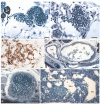
The six types of new blood vessels induced by many tumors and by Ad- vascular endothelial growth factor (VEGF)-A164 in mouse tissues. (A) Typical mother vessel (MV). (B) MVs with intraluminal extension of cytoplasmic processes, dividing the lumen into multiple smaller spaces that will eventually split to form capillaries (arrows). (C) Immunohistochemistry of a typical glomeruloid microvascular proliferation stained with antibodies against CD31. (D) Vascular malformations (VM). (E) Feeder artery. (F) Vascular mimicry (Mim) and MV in a B16 melanoma. All but (C) are Giemsa-stained 1-μm Epon sections. (A–E) were taken at early to late intervals from Ad-VEGF-A164 injection sites. Scale bars, 25 μm.
Figure 2
Figure 2
(A) Expression levels of vascular endothelial growth factor (VEGF)-A protein at successive intervals after intradermal injection of Ad-VEGF-A164. (B) Kinetics of the angiogenic and permeability responses at indicated days after Ad-VEGF-A164 injection. Ears were photographed (−EB) and then were injected intravenously with Evans blue dye for photography 30 minutes later to assess vascular permeability (+EB). Note peak of blue dye staining at day 5 when MV predominate. (C) Neovascular sites in flank skin induced by Ad-VEGF-A164. (A, B) Reprinted with permission in modified form from Nagy et al. d, day.
Figure 3
Figure 3
Schematic diagram of the angiogenic response to Ad-VEGF-A164. Reprinted with permission from Nagy et al.
Figure 4
Figure 4
Vascular basement membrane degradation as the first step in mother vessel (MV) formation. (A) Confocal microscopy of ear whole mounts from normal control (Ctl.) mice and from mice whose ears had been injected 3 days earlier with Ad-vascular endothelial growth factor (VEGF)-A164. Immediately prior to sacrifice, mice were injected intravenously with FITC-lectin (green). Ears were then immunostained with antibodies against laminin and visualized with Texas Red conjugated secondary antibodies. Note extensive loss of red laminin staining at 3 days in MV as compared with control venules. Scale bar, 50 μm. (B) Immunoblot with an antibody against laminin β1 chain performed on extracts of normal ears (time 0) and on ears harvested at 1 to 21 days following Ad-VEGF-A164 injection. Note increasing low molecular weight laminin β1 chain fragments (bracket) at 1 to 5 days. In contrast, on days 7 and 10 there is increased expression of intact laminin β1 chain (arrow), as well as high molecular weight fragments, reflecting synthesis of new vascular basement membrane (VBM) in developing glomeruloid microvascular proliferations (GMP). (C) Increased expression of proteolytically active cathepsins B, S, and L during the course of MV formation in response to Ad-VEGF-A164. NM, normal ear skin. Ad-Lac Z, control adenovirus injection. (D) Confocal microscopy of GB123 (red) and FITC-lectin (green) in normal mouse ears (NM) and in ears injected 3 days previously with Ad-VEGF-A164. Only rare stromal cells stain for GB123 (red) in NM ears, whereas pericytes, but not endothelial cells (white arrows), stain intensely red in Ad-VEGF-A164-injected ears. White asterisk indicates detaching GB123-positive pericytes. Blue color, DAPI staining. Scale bar, 20 μm. (E) Immunohistochemical staining for cystatin C in control (0) and in mouse ears 3 days after Ad-VEGF-A164 injection. Note substantial reduction in staining in MV as compared with control venules (V). Scale bar, 50 μm. (F) Reciprocal change in immunohistochemical cathepsin B and cystatin C staining in MV generated 3 days after subcutaneous injection of 106 TA3/St mammary carcinoma cells. MV exhibit reduced cysteine protease inhibitor (CPI) staining and increased cathepsin B staining as compared with control vessels (insets). Scale bar, 25 μm. Reprinted in modified form from Chang et al with permission.
Figure 5
Figure 5
Transmission electron micrographs of (A) control ear venule and (B) a mother vessel 3 days after ear injection of Ad-vascular endothelial growth factor (VEGF)-A164. (A) Typical normal venule lined by cuboidal endothelium. The cytoplasm contains prominent vesiculo-vacuolar organelles (VVOs; clear cytoplasmic vesicles) and is enveloped by a complete coating of pericytes (P). R, red blood cell. (B) Typical mother vessel (MV) is greatly enlarged sinusoid, characterized by extensive endothelial cell thinning; striking reduction in VVOs; prominent nuclei that project into the vascular lumen; mitotic figures (arrows); and decreased pericyte (P) coverage. The mother vessel lumen is packed with red blood cells, indicative of vascular leakage and resulting plasma extravasation. Inset. Reproduction of the normal venule depicted in (A) at the same magnification as the MV to illustrate differences in relative size of normal venules and mother vessels. OCUB processing. Reprinted with permission in modified form from Nagy et al. Scale bars: a, 1 μm; b, 5 μm.
Figure 6
Figure 6
Macroscopic image of a mouse ovarian tumor tumor (T) 7 days after transplant into the subcutaneous ofQ1 a syngeneic C3Heb/FeJ mouse. Angiogenesis is confined to the region indicated by the white dotted line. Note arteriovenogenesis in the vessels radiating from the tumor mass (arrows). Scale bar, 0.5 mm.

References

    1. Holash J, Maisonpierre PC, Compton D, et al. Vessel cooption, regression, and growth in tumors mediated by angiopoietins and VEGF. Science. 1999;284(5422):1994–1998. - PubMed
    1. Leenders WP, Küsters B, de Waal RM. Vessel co-option: how tumors obtain blood supply in the absence of sprouting angiogenesis. Endothelium. 2002;9(2):83–87. - PubMed
    1. Du R, Lu KV, Petritsch C, et al. HIF1alpha induces the recruitment of bone marrow-derived vascular modulatory cells to regulate tumor angiogenesis and invasion. Cancer Cell. 2008;13(3):206–220. - PMC - PubMed
    1. Folkman J. Tumor angiogenesis: therapeutic implications. N Engl J Med. 1971;285(21):1182–1186. - PubMed
    1. Warren B. The vascular morphology of tumors. In: Peterson H-I, editor. Tumor Blood Circulation: Angiogenesis, Vascular Morphology and Blood Flow of Experimental and Human Tumors. CRC Press; Boca Raton, FL: 1979. pp. 1–47.

Publication types

MeSH terms

Substances