

Porcine circovirus type 2 (PCV2) induces cell proliferation, fusion, and chemokine expression in swine monocytic cells in vitro
- PMID: 20492892
- PMCID: PMC2889373
- DOI: 10.1051/vetres/2010032
Porcine circovirus type 2 (PCV2) induces cell proliferation, fusion, and chemokine expression in swine monocytic cells in vitro
Abstract
Granulomatous lymphadenitis is one of the pathognomonic lesions in post-weaning multisystemic wasting syndrome (PMWS)-affected pigs. This unique lesion has not been reported in direct association with viral infection in pigs. The objective of the present study was to evaluate whether porcine circovirus type 2 (PCV2) alone is able to induce functional modulation in porcine monocytic cells in vitro to elucidate its possible role in the development of granulomatous inflammation. It was found that the proliferation activity of blood monocytes (Mo) and monocyte-derived macrophages (MDM) was significantly enhanced by PCV2. During monocyte-macrophage differentiation, the PCV2 antigen-containing rate and formation of multinucleated giant cells (MGC) were significantly increased in MDM when compared to those in Mo. The MDM-derived MGC displayed a significantly higher PCV2 antigen-containing rate than did the mono-nucleated MDM. Supernatants from PCV2-inoculated MDM at 24 h post-inoculation induced an increased tendency of chemotactic activity for blood Mo. At the same inoculation time period, levels of mRNA expression of the monocytic chemokines, monocyte chemoattractant protein-1 and macrophage inflammatory protein-1, also significantly increased in PCV2-inoculated MDM. The results suggest that PCV2 alone may induce cell proliferation, fusion, and chemokine expression in swine monocytic cells. Thus, PCV2 itself may play a significant role in the induction of granulomatous inflammation in PMWS-affected pigs.
© INRA, EDP Sciences, 2010.
Figures
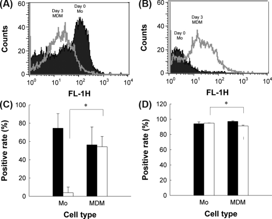


 : PCV2-inoculated; (B) ●: medium control, ▽: mock-inoculated, ■: PCV2-inoculated.
Similar articles
-
Immunopathological effects of porcine circovirus type 2 (PCV2) on swine alveolar macrophages by in vitro inoculation.Vet Immunol Immunopathol. 2006 Apr 15;110(3-4):207-19. doi: 10.1016/j.vetimm.2005.09.016. Epub 2005 Nov 28. Vet Immunol Immunopathol. 2006. PMID: 16310858
-
Experimental reproduction of postweaning multisystemic wasting syndrome in cesarean-derived, colostrum-deprived piglets inoculated with porcine circovirus type 2 (PCV2): investigation of quantitative PCV2 distribution and antibody responses.J Vet Diagn Invest. 2003 Mar;15(2):107-14. doi: 10.1177/104063870301500204. J Vet Diagn Invest. 2003. PMID: 12661720
-
Experimental transmission of porcine circovirus type 2 (PCV2) in weaned pigs: a sequential study.J Comp Pathol. 2000 Nov;123(4):258-69. doi: 10.1053/jcpa.2000.0413. J Comp Pathol. 2000. PMID: 11041995
-
Immune responses and vaccine-induced immunity against Porcine circovirus type 2.Vet Immunol Immunopathol. 2010 Aug 15;136(3-4):185-93. doi: 10.1016/j.vetimm.2010.03.025. Epub 2010 Apr 7. Vet Immunol Immunopathol. 2010. PMID: 20451259 Review.
-
Post-weaning multisystemic wasting syndrome and other PCV2-related problems in pigs: a 12-year experience.Transbound Emerg Dis. 2008 Sep;55(7):273-83. doi: 10.1111/j.1865-1682.2008.01035.x. Epub 2008 Jul 10. Transbound Emerg Dis. 2008. PMID: 18631230 Review.
Cited by
-
Identification of lncRNAs Involved in PCV2 Infection of PK-15 Cells.Pathogens. 2020 Jun 17;9(6):479. doi: 10.3390/pathogens9060479. Pathogens. 2020. PMID: 32560439 Free PMC article.
-
Porcine Circovirus Type 2 Induces Single Immunoglobulin Interleukin-1 Related Receptor (SIGIRR) Downregulation to Promote Interleukin-1β Upregulation in Porcine Alveolar Macrophage.Viruses. 2019 Nov 3;11(11):1021. doi: 10.3390/v11111021. Viruses. 2019. PMID: 31684202 Free PMC article.
-
Evaluation of the Effect of Inactivated Transmissible Gastroenteritis Virus Vaccine with Nano Silicon on the Phenotype and Function of Porcine Dendritic Cells.Viruses. 2021 Oct 26;13(11):2158. doi: 10.3390/v13112158. Viruses. 2021. PMID: 34834964 Free PMC article.
-
Transcription analysis of the porcine alveolar macrophage response to porcine circovirus type 2.BMC Genomics. 2013 May 27;14:353. doi: 10.1186/1471-2164-14-353. BMC Genomics. 2013. PMID: 23711280 Free PMC article.
-
Response of lymphocytes from pigs naturally infected with porcine respiratory disease complex at 3 different stages of development.Can J Vet Res. 2023 Apr;87(2):110-119. Can J Vet Res. 2023. PMID: 37020577 Free PMC article.
References
-
- Allan G.M., Ellis J.A., Porcine circoviruses: a review, J. Vet. Diagn. Invest. (2000) 12:3–14 - PubMed
-
- Basta S., Knoetig S.M., Spagnuolo-Weaver M., Allan G., McCullough K.C., Modulation of monocytic cell activity and virus susceptibility during differentiation into macrophages, J. Immunol. (1999) 162:3961–3969 - PubMed
-
- Borca M.V., Gudmundsdottir I., Fernandez-Sainz I.J., Holinka L.G., Risatti G.R., Patterns of cellular gene expression in swine macrophages infected with highly virulent classical swine fever virus strain Brescia, Virus Res. (2008) 138:89–96 - PubMed
-
- Byrd T.F., Multinucleated giant cell formation induced by IFN-gamma/IL-3 is associated with restriction of virulent Mycobacterium tuberculosis cell to cell invasion in human monocyte monolayers, Cell Immunol. (1998) 188:89–96 - PubMed
Publication types
MeSH terms
Substances
LinkOut - more resources
Full Text Sources
Research Materials