. 2010 Jun 11;86(6):949-56.
doi: 10.1016/j.ajhg.2010.04.012. Epub 2010 May 20.

Cranioectodermal Dysplasia, Sensenbrenner syndrome, is a ciliopathy caused by mutations in the IFT122 gene

Affiliations

Cranioectodermal Dysplasia, Sensenbrenner syndrome, is a ciliopathy caused by mutations in the IFT122 gene

Joanna Walczak-Sztulpa et al. Am J Hum Genet. .

Abstract

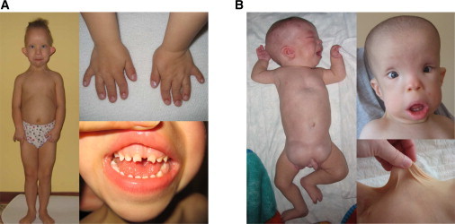
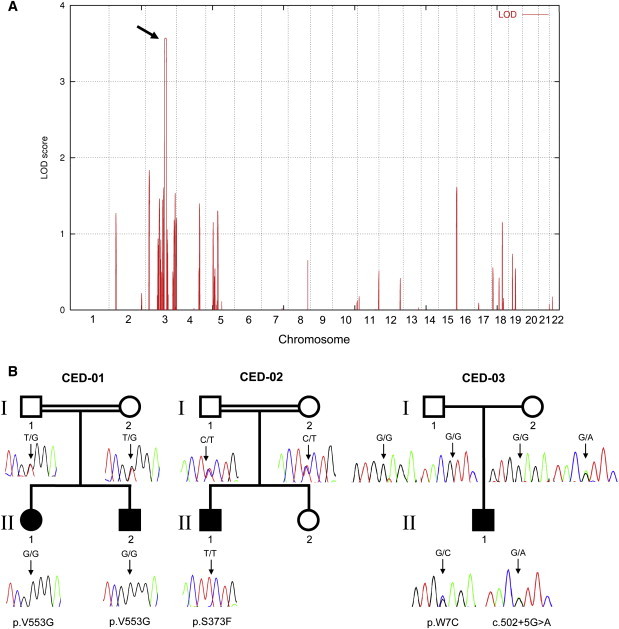
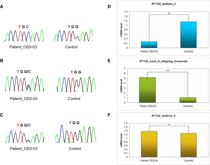
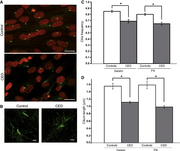
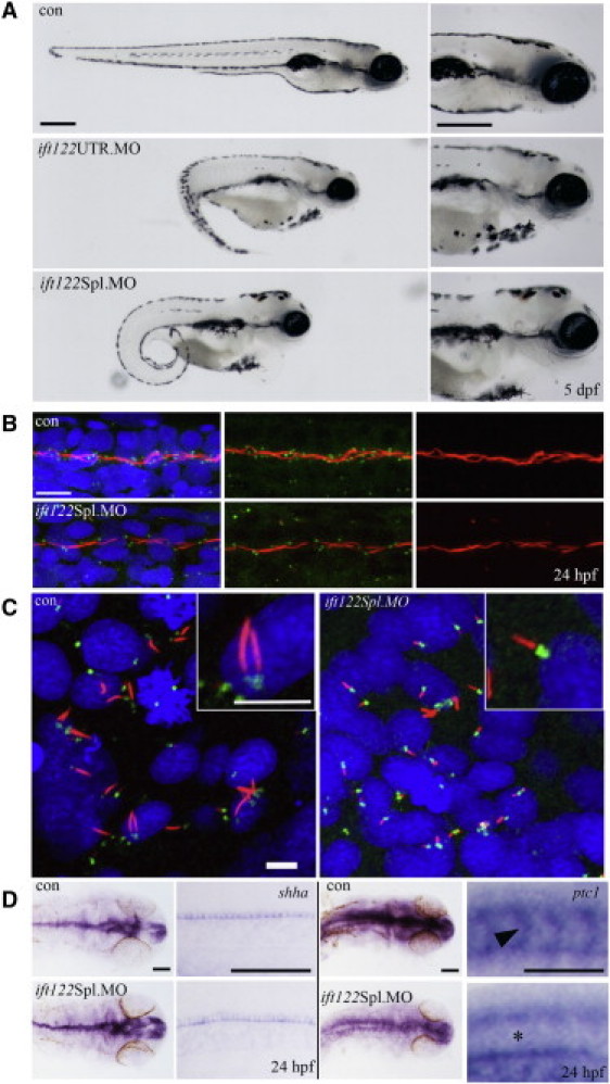
Cranioectodermal dysplasia (CED) is a disorder characterized by craniofacial, skeletal, and ectodermal abnormalities. Most cases reported to date are sporadic, but a few familial cases support an autosomal-recessive inheritance pattern. Aiming at the elucidation of the genetic basis of CED, we collected 13 patients with CED symptoms from 12 independent families. In one family with consanguineous parents two siblings were affected, permitting linkage analysis and homozygosity mapping. This revealed a single region of homozygosity with a significant LOD score (3.57) on chromosome 3q21-3q24. By sequencing candidate genes from this interval we found a homozygous missense mutation in the IFT122 (WDR10) gene that cosegregated with the disease. Examination of IFT122 in our patient cohort revealed one additional homozygous missense change in the patient from a second consanguineous family. In addition, we found compound heterozygosity for a donor splice-site change and a missense change in one sporadic patient. All mutations were absent in 340 control chromosomes. Because IFT122 plays an important role in the assembly and maintenance of eukaryotic cilia, we investigated patient fibroblasts and found significantly reduced frequency and length of primary cilia as compared to controls. Furthermore, we transiently knocked down ift122 in zebrafish embryos and observed the typical phenotype found in other models of ciliopathies. Because not all of our patients harbored mutations in IFT122, CED seems to be genetically heterogeneous. Still, by identifying CED as a ciliary disorder, our study suggests that the causative mutations in the unresolved cases most likely affect primary cilia function too.


Figures

Figure 1
Figure 1
Consanguineous CED Patients (A) CED-01, female patient 1 at the age of 4 years: note limb shortening, brachydactyly, short narrow thorax, protuberant abdomen, thin sparse hair, dolichocephaly, high prominent forehead, full cheeks, hypertelorism, small flat nose, full lower lip, prominent auricles, and small abnormally shaped teeth. (B) CED-01, male patient 2 at the age of 4 months (left pane) and 1 year (right panes): note limb shortening, pectus excavatum, bilateral large inguinal hernias, characteristic face (similar to female), and skin laxity.
Figure 2
Figure 2
Linkage Interval, Pedigrees, and IFT122 Mutations (A) Linkage interval on chromosome 3q21-3q24 (arrow) with a significant LOD score (3.57). (B) Simplified pedigrees and IFT122 mutations. CED-01: Consanguineous Polish family with two affected siblings (patients 1 and 2) homozygous for p.V553G. CED-02: Consanguineous Norwegian family (patient 3 homozygous for p.S373F). CED-03: Sporadic patient 4, who is a compound heterozygote for a splice-site mutation (c.502+5G>A) and p.W7C. Filled symbols indicate CED patient status.
Figure 3
Figure 3
Compound Heterozygosity for c.502+5G>A and p.W7C in Patient 4, CED-03 Chromatograms show sequencing results corresponding to the affected codon of p.W7C obtained with exon 6 specific primers (A) as well as primers specific for the new transcript with exon 6 skipping (B) and for isoform 4, which lacks exon 6 by default (C). Results of qPCR experiments for the corresponding amplicons show a significant (Student's t test, p < 0.01) loss of (exon 6-containing) isoform 3 (D) and a highly significant (p < 0.001) increase in expression of the transcript with exon 6 skipping (E) in RNA extracted from patient blood. Expression of isoform 4 was unaltered (F). Error bars represent the standard deviation (n = 3). Expression of the GAPDH gene was used for normalization.
Figure 4
Figure 4
Primary Cilia in Control and CED Patient Fibroblasts (A) Primary cilia from serum-starved primary fibroblasts stained with anti-acetylated-tubulin (green). Nuclei are counterstained with TO-PRO-3 (red). The scale bar represents 10 μm. (B) High magnification of cilia showing difference in axonemal lengths. The scale bar represents 1 μm. (C) Significant diffrences (p < 0.001) in average cilia frequency between control and CED patient fibroblast lines plated on gelatin and fibronectin (FN). Error bars represent the standard error of the mean (SEM). (D) Significant diffrences (p < 0.05) in average axomenal lengths between patient and control. Error bars represent the SEM.
Figure 5
Figure 5
Loss of ift122 in Zebrafish Results in a Ciliopathy-Related Phenotype (A) Gene-specific knockdown by morpholino modified antisense oligonucleotides to either the 5′UTR or to the exon 1-intron 1 splice boundary of ift122 caused reduced ocular development, melanocyte mislocalization, curly tails, renal cysts, and heart edemas by 5 dpf. All common features related to loss of ciliary genes in zebrafish. The scale bar represents 400 μm. (B) Ift122 morphants display reduced and shortened motile cilia. Immunofluorescence staining for cilia (acetylated α-tubulin, red), centrosomes (γ-tubulin, green), and nuclei (DAPI, blue) in the pronephric duct (PND) shows reduced centrosome numbers and interrupted and/or shortened cilia in morphant compared to control embryos. The scale bar represents 10 μm. (C) Cilia in the KV of ift122 Spl. Morphants are visibly shorter than uninjected controls. The scale bar represents 5 μm. (D) A mild reduction in Hh signaling was observed in morphant embryos. Embryos were analyzed for either shha or ptc1 by in situ hybridization at 24 hpf; images of the head and somites (at approximately the somite 15 position) are displayed for comparison. Shha accumulates in the floor plate of the neural tube and in the notochord (at low levels) at the 24 hpf stage; this expression remains unperturbed in morphant embryos. Ptc1 appears to be globally reduced in ift122 morphants, most notably in the somites (see asterisk) where ptc1 is normally abundant in uninjected control embryos (see arrowhead). The scale bar represents 100 μm.

References

    1. Badano J.L., Mitsuma N., Beales P.L., Katsanis N. The ciliopathies: An emerging class of human genetic disorders. Annu. Rev. Genomics Hum. Genet. 2006;7:125–148. - PubMed
    1. Lehman J.M., Michaud E.J., Schoeb T.R., Aydin-Son Y., Miller M., Yoder B.K. The Oak Ridge Polycystic Kidney mouse: Modeling ciliopathies of mice and men. Dev. Dyn. 2008;237:1960–1971. - PMC - PubMed
    1. Konstantinidou A.E., Fryssira H., Sifakis S., Karadimas C., Kaminopetros P., Agrogiannis G., Velonis S., Nikkels P.G., Patsouris E. Cranioectodermal dysplasia: A probable ciliopathy. Am. J. Med. Genet. A. 2009;149A:2206–2211. - PubMed
    1. Eke T., Woodruff G., Young I.D. A new oculorenal syndrome: Retinal dystrophy and tubulointerstitial nephropathy in cranioectodermal dysplasia. Br. J. Ophthalmol. 1996;80:490–491. - PMC - PubMed
    1. Beales P.L., Bland E., Tobin J.L., Bacchelli C., Tuysuz B., Hill J., Rix S., Pearson C.G., Kai M., Hartley J. IFT80, which encodes a conserved intraflagellar transport protein, is mutated in Jeune asphyxiating thoracic dystrophy. Nat. Genet. 2007;39:727–729. - PubMed

Publication types