. 2010 May 18;18(5):828-40.
doi: 10.1016/j.devcel.2010.03.016.

Phosphatidylinositol 4-phosphate controls both membrane recruitment and a regulatory switch of the Rab GEF Sec2p

Affiliations

Phosphatidylinositol 4-phosphate controls both membrane recruitment and a regulatory switch of the Rab GEF Sec2p

Emi Mizuno-Yamasaki et al. Dev Cell. .

Abstract

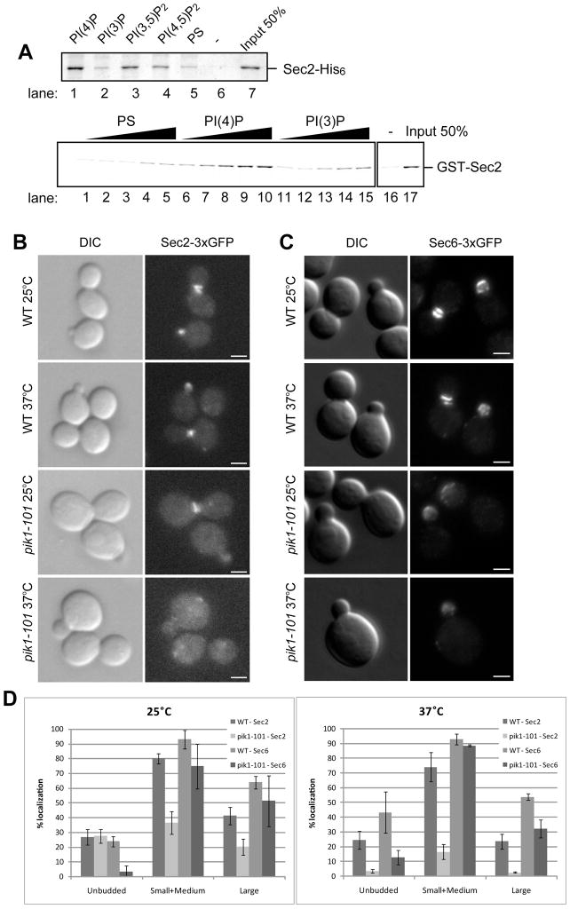
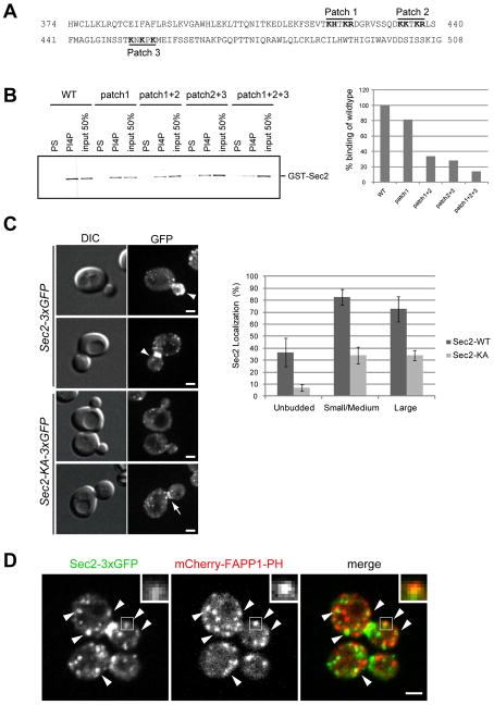
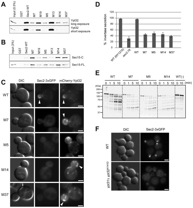
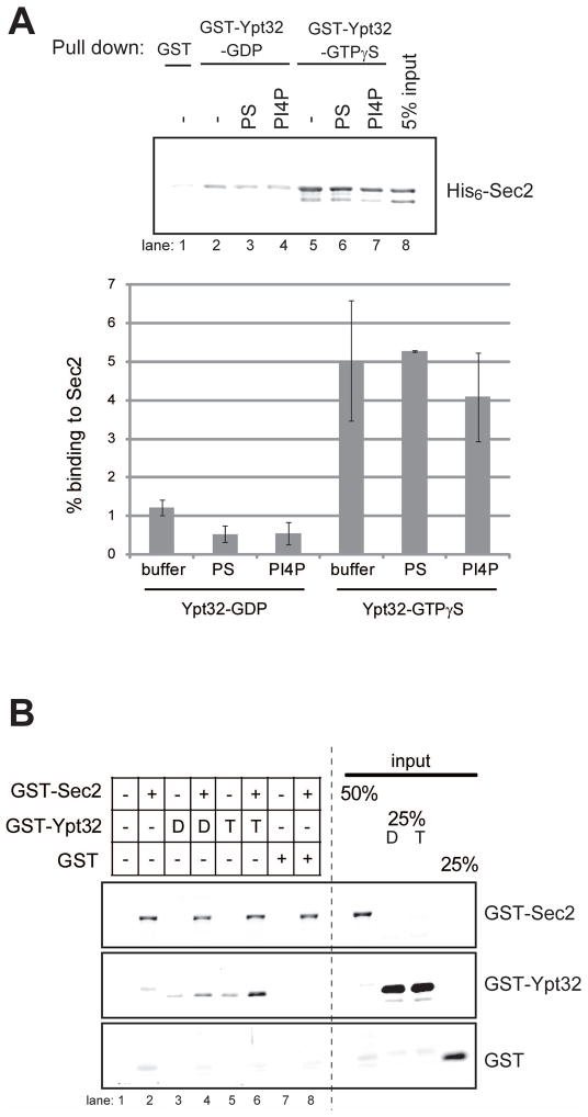
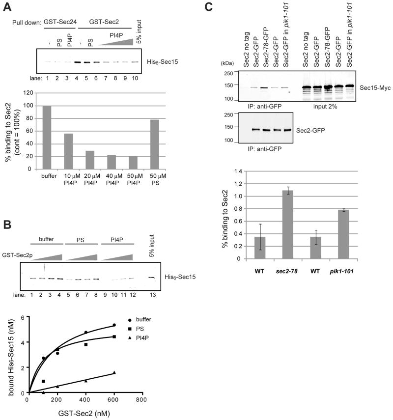
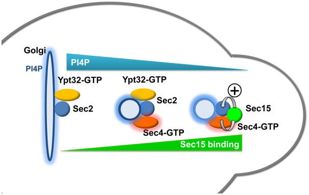
Sec2p is the guanine nucleotide exchange factor (GEF) that activates the Rab GTPase Sec4p on secretory vesicles. Sec2p also binds a Rab acting earlier in the secretory pathway, Ypt32-GTP, forming a Rab GEF cascade. Ypt32p and the Sec4p effector Sec15p (a component of the exocyst complex) compete for binding to Sec2p. Indeed Ypt32p initially recruits Sec2p, but subsequently allows a handoff of active Sec2p/Sec4p to Sec15p. Intriguingly, Golgi-associated phosphatidylinositol 4-phosphate (PI4P) works together with Ypt32-GTP in this context. PI4P inhibits Sec2p-Sec15p interactions, promoting recruitment of Sec2p by Ypt32p as secretory vesicles form. However, PI4P levels appear to decline as vesicles reach secretory sites, allowing Sec15p to replace Ypt32p as vesicles mature. In this way, the regulation of PI4P levels may switch Sec2p/Sec4p function during vesicle maturation, from a Rab GEF recruitment cascade involving Ypt32p to an effector positive feedback loop involving Sec15p.


Figures

Figure 1
Figure 1
(A) Sec2p directly binds to Phosphoinsitides. (top panel) Sec2p-His6 (120 nM) purified from E. Coli was incubated by itself (lane 6) or with 0.5 mM liposomes containing 50/30/15 mol% of PC/PE/PS and 5 mol% of the indicated phosphoinositides (lanes 1–4), or 45/30/25 mol% of PC/PE/PS (lane 5). Liposomes were precipitated by centrifugation at 100,000 × g and bound proteins were detected by Coomassie blue staining. (bottom panel) GST-Sec2p purified from E. Coli was incubated with 0.05, 0.1, 0.25, 0.375, and 0.5 mM of liposomes containing 10 mol% of PS, PI4P, or PI3P. Liposomes were precipitated by centrifugation at 100,000 x g and bound proteins were detected with anti-Sec2p antibody. (B) Sec2p is mislocalized in pik1-101 cells. Localization of Sec2-3xGFP (B) and Sec6-3xGFP (C) was examined in wildtype (WT) and pik1-101 cells. Cells were grown overnight at 25°C in a synthetic medium containing 2% glucose and then shifted to 37°C for 60 minutes. Cells were immediately fixed and examined as described in Experimental procedures. Bars are 2 μm. (D) Cells were classified into three categories (unbudded, small/medium buds, or large buds). A total of approximately 200 cells were counted for each experiment. Values indicate the percentage of cells showing bud or mother-daughter neck localization of Sec2-3xGFP or Sec6-3xGFP. Mean and S.D. of three different experiments are shown.
Figure 2
Figure 2. Sec2p-PI4P interaction is required for Sec2p localization
(A) The Sec2 sequence between a.a. 374 and 508 is necessary for PI4P binding. Positively charged patches are shown in bold. (B) GST-tagged Sec2 wild-type (WT), or Sec2 with mutations in positively charged patches 1, 2, or 3, were purified from E. Coli. Proteins were incubated with liposomes containing 10 mol% PS or PI4P. Liposomes were precipitated by centrifugation at 100,000 x g and bound proteins were detected with anti-Sec2 antibody. The intensity of the bands was quantified using Image J. (C) Localization of Sec2-3xGFP or Sec2-KA-3xGFP was examined. Cells were grown overnight at 25°C in a synthetic medium containing 2% glucose. Bars are 2 μm. Arrowheads indicate normal localization of wildtype Sec2. Arrow in bottom panel shows normal localization of Sec2-KA, but with reduced intensity. A total of approximately 80 cells were counted for each experiment. Values indicate the percentage of cells showing bud or mother-daughter neck localization of Sec2-3xGFP. Mean and S.D. of three different experiments are shown. (D) The localization of Sec2-3xGFP (left), mCherry-FAPP1-PH (middle) and the merge (right) was examined in wildtype cells. Cells were grown at 25°C in a synthetic medium containing 2% glucose and then live cells were analyzed with a spinning disk confocal microscope. Insets and arrowheads show colocalization between Sec2-3xGFP and mCherry-FAPP1-PH. Bar is 2 μm.
Figure 3
Figure 3. Overexpression of Sec2p, Ypt32p and Sec4p partially suppresses the growth defect of pik1-101
(A) Wild-type (WT), pik1-101, and pik1-101 cells overexpressing Sec2p, Ypt1p, Ypt32p, or Sec4p from a high-copy number, 2μ-based plasmid were spotted onto YPD plates in 5-fold serial dilutions. Cells were grown at the indicated temperatures for two days. Two independent transformants were analyzed for each plasmid. (B) Localization of Sec2-3xGFP was examined in wildtype (WT, top panels), WT harboring the Ypt32 overexpressing vector (second panels from top), pik1-101 (third panels from top), or pik1-101 harboring the Ypt32 overexpressing vector (bottom panels). Cells were grown at 25°C overnight in a synthetic medium containing 2% glucose and then shifted to 37°C for 60 minutes. Cells were immediately fixed and examined. Images shown are cells after the 37°C shift. Bars are 2μm. (C) Cells were classified into three categories (unbudded, small/medium buds, or large buds). A total of approximately 100 cells were counted for each experiment. Values indicate the percentage of cells showing bud or mother-daughter neck localization of Sec2-3xGFP. (D) Tetrads from a diploid carrying the sec2-KA, ypt31Δ and ypt32-A141D alleles. ypt31Δ ypt32-A141D spores are shown within circles. ypt31Δ ypt32-A141D sec2-KA triple mutants are shown within squares.
Figure 4
Figure 4. Sec2p-Ypt32p interaction is required for Sec2p localization, growth, and secretion
(A) An in vitro-binding assay confirms the loss of the Ypt32p-Sec2p interaction in Sec2 mutants. GST-Sec2p and His6-Ypt32p were purified from E. coli. Ypt32p was eluted, preloaded with GTPγS, and used in binding reactions with Sec2p and Sec2p mutants immobilized on beads, as described in Experimental procedures. Bound proteins were detected with anti-Ypt32 antibody. (B) In vitro binding of Sec15p is increased for Sec2 mutants that show decreased binding to Ypt32p. GST-Sec2p and His6-Sec15p (Sec15-FL) or His6-Sec15p(559–901) (Sec15-C) were purified from E. coli. Sec15p was eluted and incubated with GST-Sec2p immobilized on glutathione beads. Bound proteins were detected with anti-Sec15 antibody. (C) Sec2 mutants are mislocalized. The localization of Sec2p-3xGFP and mCherry-Ypt32p were examined in wildtype (WT) and various sec2 mutants. Cells were grown overnight at 25°C in synthetic medium containing 2% glucose and live cells were observed. Arrowheads in WT indicate the bud tip localization of Sec2p and Ypt32p. Bars are 2 μm. (D) Secretion of invertase is blocked in sec2 mutants. The percentage of the secreted invertase was examined in untransformed wild type cells (NY1210), sec2-78, wild type expressing Sec2-3xGFP, and the four M-mutants fused to 3xGFP. Cells were grown overnight at 25°C in YP medium with 2% glucose. 1 OD unit of cells was resuspended in YP medium containing 0.1% glucose and cultured at 37°C for 60 min. The amount of invertase secreted during this time period was measured as described in Experimental procedures. The mean and S.D of three different experiment are shown. (E) Ypt32p-binding deficient sec2 mutant shows different conformation. GST-tagged Sec2 and various Sec2 mutant proteins were purified from yeast and the purified proteins were then incubated with or without (−) trypsin. Aliquots were withdrawn from reaction tubes at 0 min, 1 min, 5 min, and 10 min. Proteolytic fragments were detected with an anti-Sec2p antibody that recognizes the N-terminal part of Sec2p. (F) Sec2 was mislocalized in ypt31Δ ypt32A141D cells. Localization of Sec2-3xGFP was examined in wildtype (top panels) and ypt31Δ ypt32A141D (bottom panels) cells. Cells were grown overnight at 25°C in synthetic medium containing 2% glucose. Cells were fixed and examined. Bars are 2 μm.
Figure 5
Figure 5. Three-way interaction between Sec2p, PI4P and Ypt32-GTP
(A) PI4P does not significantly affect Ypt32-binding to Sec2. GST or GST-Ypt32p (0.60 μM) were immobilized on glutathione-Sepharose beads and preloaded with GDP or GTPγS. His6-Sec2p (0.11 μM) was incubated with buffer (−) or liposome containing 10 mol% (50 μM) of PS or PI4P for 30 min. Then the Sec2-liposome complex was added to the Ypt32-conjugated beads and incubated for an additional 60 min. After the reaction, beads were washed with buffer containing 0.05% Triton X-100. Bound proteins were detected with anti-Ypt32p antibody. The intensity of the bands was quantified using Image J. The percentage of bound Sec2p was calculated and indicated. The mean and S.D of three different experiments are shown. (B) PI4P and Ypt32p can bind Sec2p at the same time. GST-Sec2p, GST-Ypt32p or GST were purified from E. Coli and eluted from glutathione-Sepharose beads. GST-Ypt32p was preloaded with GDP (D) or GTPγS (T). GST-Sec2p (0.15 μM), nucleotide-loaded GST-Ypt32p (0.60 μM), or GST (0.60 μM) were incubated with liposome containing 10 mol% PI4P for 30 min. Liposomes were precipitated by centrifugation at 100,000 × g and bound proteins were analyzed with anti-GST antibody. Lane 6 shows increased association of Ypt32-GTP with PI4P liposomes in the presence of Sec2p, suggesting the formation of a ternary complex.
Figure 6
Figure 6. PI4P inhibits binding of Sec15p to Sec2p
(A) PI4P inhibits the binding of Sec15p to Sec2p. GST-Sec24p or GST-Sec2p (0.10 μM) were immobilized on glutathione-Sepharose beads and preincubated with buffer (−) or 0.5 mM liposome containing 50 μM of PS, or 10 to 50 μM of PI4P for 30 min. Then 50 nM of purified His6-Sec15p was added to the beads and incubated for an additional 60 min. After the reaction, beads were washed with buffer containing 0.05% Triton X-100. Bound Sec15p was detected with anti-Sec15p antibody. The intensity of the bands was quantified using Image J. (B) 100 to 600 nM of GST-Sec2p was immobilized on glutathione-Sepharose beads and preincubated with buffer (−) or 0.5 mM liposome containing 50 μM of PS, or 50 μM of PI4P for 30 min. Then 50 nM of purified His6-Sec15p was added to the beads and incubated for an additional 60 min. After the reaction, beads were washed with buffer containing 0.05% Triton X-100. Bound proteins were detected with anti-Sec15p antibody. The intensity of the bands was quantified using Image J. Results shown here are representative of three independent experiments. (C) The Sec2p-Sec15p interaction is enhanced in pik1-101 cells. Sec2p-GFP was immunoprecipitated with anti-GFP antibody from wild-type or pik1-101 cell lysates. Sec2-78p-GFP was immunoprecipitated from the sec2-78 mutant. As a negative control, cells expressing untagged Sec2p were used. Co-precipitated Sec15p-13xMyc was detected with anti-Myc antibody (top panel). Sec2-GFP in the immunoprecipitates was detected by immunoblotting with anti-GFP antibody (bottom panel). The intensity of the bands was quantified using Image J. The percentage of Sec15p in the immunoprecipitate was calculated and indicated. The mean and S.D of three different experiment are shown.
Figure 7
Figure 7. A model for recruitment and regulation of Sec2p by PI4P
From left to right: Sec2p is initially recruited to the Golgi membrane through the combined signals from PI4P and Ypt32-GTP. The interaction of PI4P with Sec2p also serves to block the binding of Sec15p with Sec2p, thereby facilitating the interaction of Sec2p with Ypt32-GTP. Vesicles bud off of the Golgi carrying Sec2p and Ypt32-GTP, but containing reduced levels of PI4P. Sec2p activates Sec4p, which then recruits its effector, Sec15p. The reduced level of PI4P on the vesicle membrane allows Sec15p to competitively replace Ypt32-GTP on Sec2p. This generates a positive feedback loop in which Sec2p activates Sec4p, Sec4-GTP recruits Sec15p and Sec15p helps retains Sec2p on the vesicle. The elevated levels of Sec4-GTP and Sec15p prepare the vesicle to tether, dock and fuse with the plasma membrane.

Similar articles

Cited by

References

    1. Audhya A, Emr SD. Stt4 PI 4-kinase localizes to the plasma membrane and functions in the Pkc1-mediated MAP kinase cascade. Developmental cell. 2002;2:593–605. - PubMed
    1. Audhya A, Foti M, Emr SD. Distinct roles for the yeast phosphatidylinositol 4-kinases, Stt4p and Pik1p, in secretion, cell growth, and organelle membrane dynamics. Molecular biology of the cell. 2000;11:2673–2689. - PMC - PubMed
    1. Benli M, Doring F, Robinson DG, Yang X, Gallwitz D. Two GTPase isoforms, Ypt31p and Ypt32p, are essential for Golgi function in yeast. The EMBO journal. 1996;15:6460–6475. - PMC - PubMed
    1. Boyd C, Hughes T, Pypaert M, Novick P. Vesicles carry most exocyst subunits to exocytic sites marked by the remaining two subunits, Sec3p and Exo70p. The Journal of cell biology. 2004;167:889–901. - PMC - PubMed
    1. Christoforidis S, McBride HM, Burgoyne RD, Zerial M. The Rab5 effector EEA1 is a core component of endosome docking. Nature. 1999a;397:621–625. - PubMed

Publication types

MeSH terms

Substances