. 2010 May 18;18(5):841-8.
doi: 10.1016/j.devcel.2010.04.007.

Conformation switching of clathrin light chain regulates clathrin lattice assembly

Affiliations

Conformation switching of clathrin light chain regulates clathrin lattice assembly

Jeremy D Wilbur et al. Dev Cell. .

Abstract

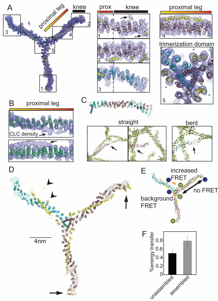
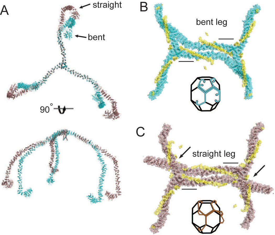
Clathrin-coated vesicle formation is responsible for membrane traffic to and from the endocytic pathway during receptor-mediated endocytosis and organelle biogenesis, influencing how cells relate to their environment. Generating these vesicles involves self-assembly of clathrin molecules into a latticed coat on membranes that recruits receptors and organizes protein machinery necessary for budding. Here we define a molecular mechanism regulating clathrin lattice formation by obtaining structural information from co-crystals of clathrin subunits. Low resolution X-ray diffraction data (7.9-9.0 A) was analyzed using a combination of molecular replacement with an energy-minimized model and noncrystallographic symmetry averaging. Resulting topological information revealed two conformations of the regulatory clathrin light chain bound to clathrin heavy chain. Based on protein domain positions, mutagenesis, and biochemical assays, we identify an electrostatic interaction between the clathrin subunits that allows the observed conformational variation in clathrin light chains to alter the conformation of the clathrin heavy chain and thereby regulates assembly.


Figures

Figure 1
Figure 1. 7.9 Å resolution electron density of the clathrin hub-LCb complex and conformational variability of clathrin light chains
(A) Model of the hub-LCb complex determined by X-ray crystallography with 2Fo-Fc electron density (0.06e/A3, 1.2σ, blue mesh). Details are shown in numbered boxes indicating the regions. Regions 1 and 2 show CHC straight knee conformations (brown ribbons) and region 3 shows the CHC bent knee conformation (blue ribbons), N-terminal to the proximal (prox) leg. CLC residues are shown as yellow ribbons and highlighted by arrows in region 1. Asterisks in region 5 (trimerization domain) indicate helices from the C-terminus of CLC that make contacts with two different CHCs. (B) The high resolution structure of the clathrin heavy chain proximal leg (1B89, green ribbon, Ybe et al., 1999) fitted into the 2Fo-Fc electron density from the proximal leg region. Arrows point to density corresponding to CLC. (C) Straight (brown ribbons) or bent (blue ribbons) legs from the 7.9 Å, spacegroup I4122 structure were aligned with each other in PYMOL. Boxes show contacts between crystal symmetry-mates (green) surrounding the N-terminus of straight or bent legs, colored as above. Arrows point to areas of symmetry-mate contact closest to the N-terminus of each leg. (D) Structural model of the clathrin hub-LCb complex. CHC helices are in brown (straight conformation) and blue (bent conformation) and CLC is in yellow. CLC extends from the trimerization domain to the knee region on two legs with straight knees (arrows) while on the bent leg CLC forms a more compact structure in the N-terminal region (arrowheads). (E) Diagram indicating the possible FRET interactions and the locations of the fluorescent dyes in blue and green. Orange arrows indicate distances favorable for FRET between two clathrin light chains (background at the trimerization domain or between termini of clathrin light chains). Dashed black arrow indicates a distance unfavorable for FRET. (F) CLC labeled randomly with two different fluorophores was bound to clathrin hub and these complexes were mixed with complexes of hub with unlabeled CLC to prevent intermolecular energy transfer (as determined in Figure S1D). FRET was measured in either the unassembled or the assembled state. Each bar represents the mean ±SEM (n=3).
Figure 2
Figure 2. The KR loop in the clathrin heavy chain knee participates in electrostatic regulation of assembly by clathrin light chain
(A) Expanded boxed region shows the position of the KR loop (blue spheres, residues 1161–1165 in CHC) relative to CLC. The straight knee conformation (brown) associated with the extended form of CLC (yellow) and the bent knee conformation (cyan) associated with the compact form of CLC (not shown) are overlaid. The KR loop is indicated by red arrowheads. Major deviations between the overlaid structures occur distal to the KR loop. (B) Expanded box region of a, rotated 90° (straight knee only). (C) Alignment of various CHC protein sequences around the KR loop (bold, yellow highlight). Red highlights show differences that alter the charge distribution. Residues mutated to alter the KR loop charge indicated by asterisks (K1163E and R1165D). (D) Surface plasmon resonance (SPR) analysis of clathrin light chain interactions with wild type clathrin hub or KR loop mutant (mut) hub. Human neuronal LCb was immobilized on the SPR chip surface and wild type or KR loop mutant hub flowed over at different concentrations. The fitted binding affinity for wild type was 25 nM while KR loop mutant was 59.1 nM. The difference in affinity for CLC was due to a slowed on-rate for the KR loop mutant likely due to decreased electrostatic attraction. (E) Purified hubs (wild-type, WT or mutant, KR) were saturated with CLC (wild type neuronal LCb (WT CLC) or neuronal LCb with E20ED22 mutated to K20KK22 (MUT CLC)) or left unoccupied and assayed for assembly at pH 6.7 by light scattering. The ratio of assembly signal for hub with light chain to assembly signal for hub without light chain for is plotted for each reaction (mean±SEM). (F) Representative kinetic assembly data for WT or KR loop mutant (MUT) hub with or without CLC bound, as indicated. Assembly is detected photometrically by an increase in light scatter at OD320.
Figure 3
Figure 3. Modeling clathrin light chain-induced conformational changes in the triskelion knee and comparison to the assembled clathrin lattice
(A) Clathrin heavy chain triskelia with all straight knees or all bent knees were modeled by alignment of distal legs (residues 1–1130) from the cryoEM structure of Fotin et al. (2004) with the hub structures determined here. Straight conformation (brown) and bent conformation (cyan) of knee regions are shifted ~30° starting from the KR loop hinge point. (B and C) Alignment of two modeled hubs, both with a single full length bent knee leg (B) or straight knee leg (C), with two assembled triskelia in the cryoEM structure (Fotin et al., 2004)(1XI4). Black bars indicate regions used to align cryoEM and hub crystal structures and the clathrin light chains are shown in yellow in their relative positions in the hub-LCb complex. Arrows indicated point of steric clashes in C. Insets indicate the geometric alignment of the bent or straight leg triskelia as modeled in (A) along edges of the clathrin basket.

Similar articles

Cited by

References

    1. Brodsky FM, Chen CY, Knuehl C, Towler MC, Wakeham DE. Biological basket weaving: formation and function of clathrin-coated vesicles. Annu Rev Cell Dev Biol. 2001;17:517–568. - PubMed
    1. Brunger AT. Version 1.2 of the Crystallography and NMR system. Nat Protoc. 2007;2:2728–2733. - PubMed
    1. Brunger AT, Adams PD, Clore GM, DeLano WL, Gros P, Grosse-Kunstleve RW, Jiang JS, Kuszewski J, Nilges M, Pannu NS, et al. Crystallography & NMR system: A new software suite for macromolecular structure determination. Acta Crystallogr D Biol Crystallogr. 1998;54:905–921. - PubMed
    1. Chen CY, Brodsky FM. Huntingtin-interacting protein 1 (Hip1) and Hip1-related protein (Hip1R) bind the conserved sequence of clathrin light chains and thereby influence clathrin assembly in vitro and actin distribution in vivo. J Biol Chem. 2005;280:6109–6117. - PubMed
    1. Chen CY, Reese ML, Hwang PK, Ota N, Agard D, Brodsky FM. Clathrin light and heavy chain interface: alpha-helix binding superhelix loops via critical tryptophans. Embo J. 2002;21:6072–6082. - PMC - PubMed

Publication types

MeSH terms