. 2010 Sep 10;584(17):3831-8.
doi: 10.1016/j.febslet.2010.05.019. Epub 2010 May 21.

Dyskeratosis congenita

Affiliations

Dyskeratosis congenita

Monica Bessler et al. FEBS Lett. .

Abstract

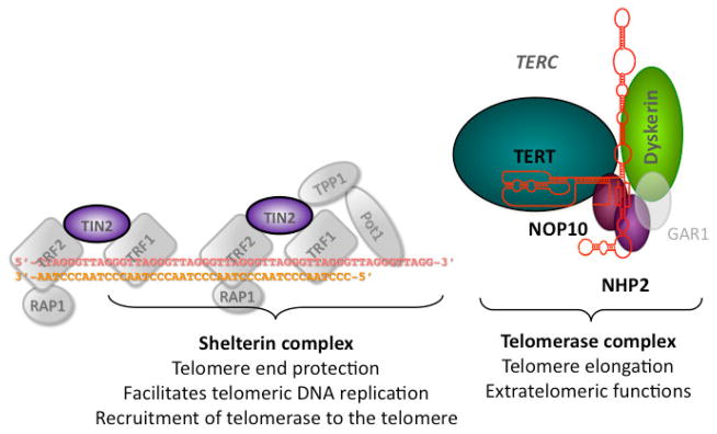
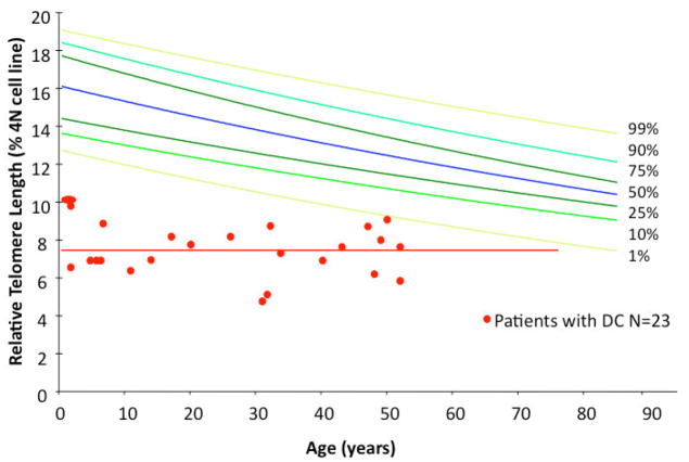
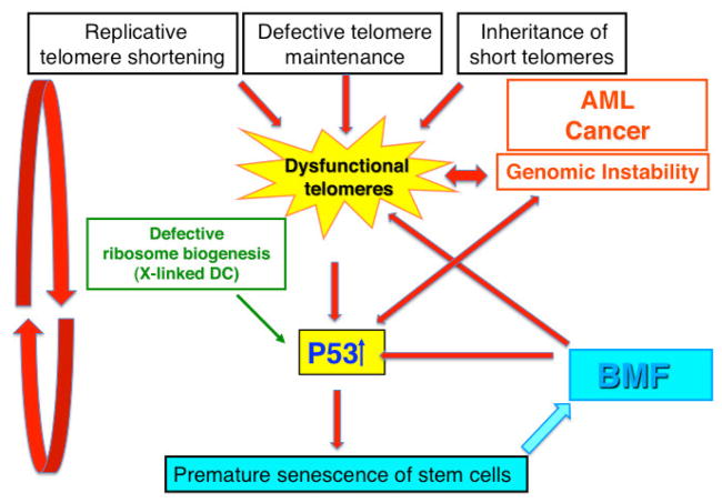
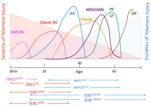
Dyskeratosis congenita (DC) was originally defined as a rare inherited bone marrow failure (BMF) syndrome associated with distinct mucocutaneous features. Today DC is defined by its pathogenetic mechanism and mutations in components of the telomere maintenance machinery resulting in excessively short telomeres in highly proliferating tissues. With this new definition the disease spectrum has broadened and ranges from intrauterine growth retardation, cerebellar hypoplasia, and death in early childhood to asymptomatic mutation carriers whose descendants are predisposed to malignancy, BMF, or pulmonary disease. The degree of telomere dysfunction is the major determinant of disease onset and manifestations.


Figures

Figure 1
Figure 1
Diagram of the telomere ends and of telomerase and the components mutated in patients with Dyskeratosis Congenita. The 6 proteins of the shelterin complex (TRF1, TRF2, RAP1, TIN2, TPP1, and POT1) protect the telomere from being recognized as a double strand break and regulate the access of telomerase. Telomerase is a ribonucleoprotein consisting of the catalytic subunit (TERT), its RNA component TERC, and the four H/ACA RNA associated proteins dyskerin, NHP2, NOP10 and GAR1. The 6 genes known to be mutated in patients with DC are highlighted in color. No mutations have been indentified in GAR1, or the other components of the shelterin complex.
Figure 2
Figure 2
Telomere length distribution in normal controls and patients with BMF due to DC. The colored lines represent the 1st, 5th, 25th, 50th, 75th, 95th and 99th percentiles of telomere length determined in peripheral blood mononuclear cells in 250 normal controls between the ages of 1 and 94 years old. In red are the telomere lengths in peripheral blood mononuclear cells isolated from patients with BMF and DC in whom a mutation in a DC associated gene was confirmed by genetic testing.
Figure 3
Figure 3
Model of the pathogenesis of BMF and malignancy in patients with DC. Our hypothesis is that dysfunctional telomeres play a central role in the pathogenesis of BMF and malignancy in patients with DC. Dysfunctional telomeres cause the activation of the p53 pathway leading to cell cycle arrest, senescence, and cell death in replicating progenitor cells. Depletion of proliferating progenitor cells recruits new stem cells into cell cycle, thus initiating a vicious cycle that ultimately leads to the depletion of stem cells and to the clinical picture of AA. Increased oxidative stress in the environment of BMF and defective ribosome biogenesis in the X-linked and some rare autosomal recessive forms of DC may additionally contribute to the activation of the p53 pathway. Dysfunctional telomeres cause genomic instability that leads to cancer predisposition in patients with DC.
Figure 4
Figure 4
Age-related variable disease expression of mutations in DC associated genes. The clinical phenotype of mutations in DC related genes varies with the age of onset. Mutations that most severely compromise telomere maintenance manifest early in life leading to the more severe forms of DC including the HHS (Hoyeraal Hreidarrson syndrome) or RS (Revesz syndrome) They may be caused by mutations in the DKC1 and TINF2 gene. Classic DC is most frequently caused by DCK1 gene mutations or homozygous or compound heterozygous mutations in the TERC or TERT genes. Heterozygous mutations in TERC or TERT genes may be associated with BMF, whereas mucco-cutaneous features may be mild or missing (aplastic anemia, AA). Pulmonary fibrosis (PF) or liver fibrosis (LF) are DC manifestations that clinically become obvious later in life. They may present as the only clinical feature in patients heterozygous for TERT or TERC gene mutations. Malignancy (cancer and MDS/AML) is most frequently seen in patients with mild-moderate disease severity and occurs most frequently in the third, fourth and fifth decade. In patients with TERT or TERC gene mutations, MDS or MDS/AML might be the initial presentation at diagnosis. The severity and the duration of dysfunctional telomere maintenance are important factors that determine the disease pathology. M=mutant.

References

    1. Bessler M, Mason P, Link D, Wilson D. Nathan and Oski’s Hematology of Infancy and Childhood. In: Orkin S, Nathan D, Ginsburg D, Look A, Fisher D, Lux S, editors. Saunders; Philadelphia: 2008. pp. 307–395.
    1. de Lange T. How telomeres solve the end-protection problem. Science. 2009;326:948–52. - PMC - PubMed
    1. O’Sullivan RJ, Karlseder J. Telomeres: protecting chromosomes against genome instability. Nat Rev Mol Cell Biol. 2010;11:171–81. - PMC - PubMed
    1. de Lange T, Shiue L, Myers RM, Cox DR, Naylor SL, Killery AM, Varmus HE. Structure and variability of human chromosome ends. Mol Cell Biol. 1990;10:518–27. - PMC - PubMed
    1. Blackburn EH. Structure and function of telomeres. Nature. 1991;350:569–73. - PubMed

Publication types

MeSH terms