

Corpus callosum abnormalities and their association with psychotic symptoms in patients with schizophrenia
- PMID: 20494336
- PMCID: PMC2900500
- DOI: 10.1016/j.biopsych.2010.03.025
Corpus callosum abnormalities and their association with psychotic symptoms in patients with schizophrenia
Abstract
Background: While the neuroanatomical underpinnings of the functional brain disconnectivity observed in patients with schizophrenia (SZ) remain elusive, white matter fiber bundles of the brain are a likely candidate, given that they represent the infrastructure for long-distance neural communication.
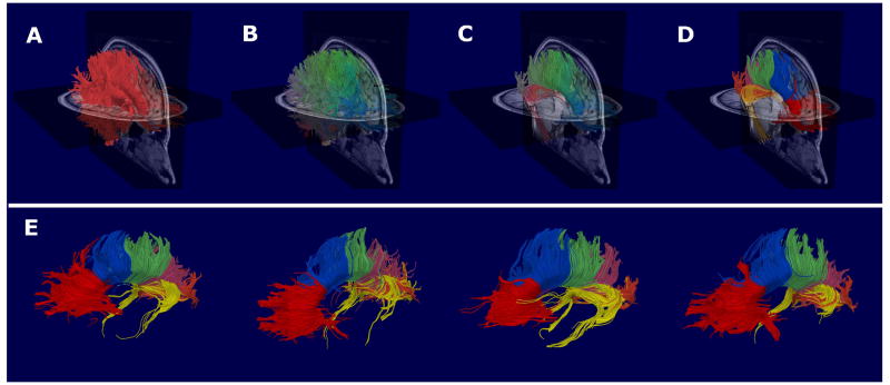
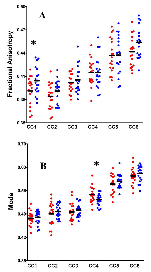
Methods: This study investigated for diffusion abnormalities in 19 patients with chronic SZ, relative to 19 matched control subjects, across tractography-defined segments of the corpus callosum. Diffusion-weighted images were acquired with 51 noncollinear gradients on a 3T scanner (1.7 mm isotropic voxels). The corpus callosum was extracted by means of whole-brain tractography and automated fiber clustering and was parcelled into six segments on the basis of fiber trajectories. The diffusion indexes of fractional anisotropy (FA) and mode were calculated for each segment.
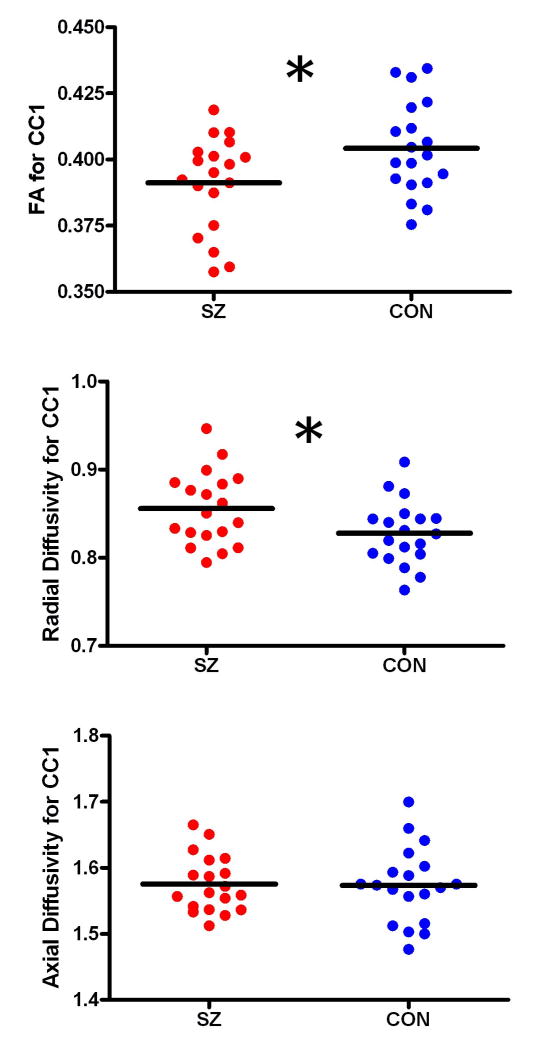
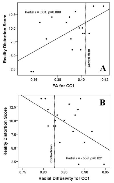
Results: Relative to the healthy control subjects, the SZ patients exhibited mode increases in the parietal fibers, suggesting a relative absence of crossing fibers. Schizophrenia patients also exhibited FA reductions in the frontal fibers, which were underpinned by increases in radial diffusivity, consistent with myelin abnormalities. Significant correlations were observed between patients' degree of reality distortion and their FA and radial diffusivity, such that the most severely psychotic patients were the least abnormal in terms of their frontal fiber diffusivity.
Conclusions: The SZ patients exhibited a variety of diffusion abnormalities in the corpus callosum, which were related to the severity of their psychotic symptoms. To the extent that diffusion abnormalities influence axonal transmission velocities, these results provide support for those theories that emphasize neural timing abnormalities in the etiology of schizophrenia.
Copyright 2010 Society of Biological Psychiatry. Published by Elsevier Inc. All rights reserved.
Conflict of interest statement
Figures
References
-
- Andreasen NC. A unitary model of schizophrenia: Bleuler's “fragmented phrene” as schizencephaly. Archives of General Psychiatry. 1999;56:781–787. - PubMed
-
- Friston K. Schizophrenia and the disconnection hypothesis. Acta Psychiatr Scand Suppl. 1999;395:68–79. - PubMed
-
- Bartzokis G. Schizophrenia: breakdown in the well-regulated lifelong process of brain development and maturation. Neuropsychopharmacology. 2002;27:672–683. - PubMed
-
- Norman RM, Malla AK, Williamson PC, Morrison-Stewart SL, Helmes E, Cortese L. EEG coherence and syndromes in schizophrenia. Br J Psychiatry. 1997;170:411–415. - PubMed
-
- Fletcher P, McKenna PJ, Friston KJ, Frith CD, Dolan RJ. Abnormal cingulate modulation of fronto-temporal connectivity in schizophrenia. Neuroimage. 1999;9:337–342. - PubMed
Publication types
MeSH terms
Grants and funding
- R01 MH040799/MH/NIMH NIH HHS/United States
- CA089017-06A2/CA/NCI NIH HHS/United States
- T32 MH 016259/MH/NIMH NIH HHS/United States
- R01 MH 50747/MH/NIMH NIH HHS/United States
- R25 CA089017/CA/NCI NIH HHS/United States
- K05 MH 070047/MH/NIMH NIH HHS/United States
- MH 040799/MH/NIMH NIH HHS/United States
- P50 MH 080272/MH/NIMH NIH HHS/United States
- R03 MH068464-0/MH/NIMH NIH HHS/United States
- T32 MH016259/MH/NIMH NIH HHS/United States
- R03 MH068464/MH/NIMH NIH HHS/United States
- K05 MH070047/MH/NIMH NIH HHS/United States
- R01 MH050740/MH/NIMH NIH HHS/United States
- U54 EB005149/EB/NIBIB NIH HHS/United States
- P50 MH080272/MH/NIMH NIH HHS/United States
LinkOut - more resources
Full Text Sources
Medical
