. 2010 Oct 28;157(1-2):81-90.
doi: 10.1016/j.autneu.2010.03.022. Epub 2010 May 21.

Acupuncture, the limbic system, and the anticorrelated networks of the brain

Affiliations

Acupuncture, the limbic system, and the anticorrelated networks of the brain

Kathleen K S Hui et al. Auton Neurosci. .

Abstract

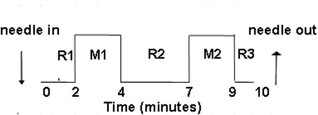
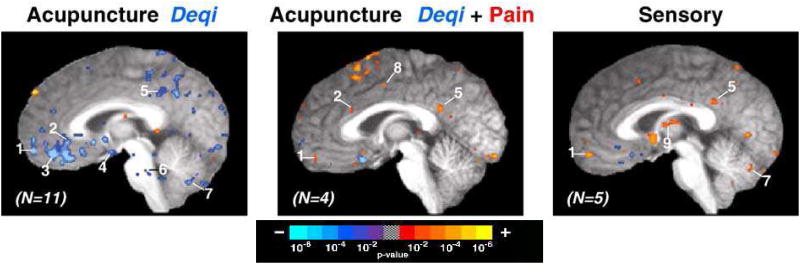
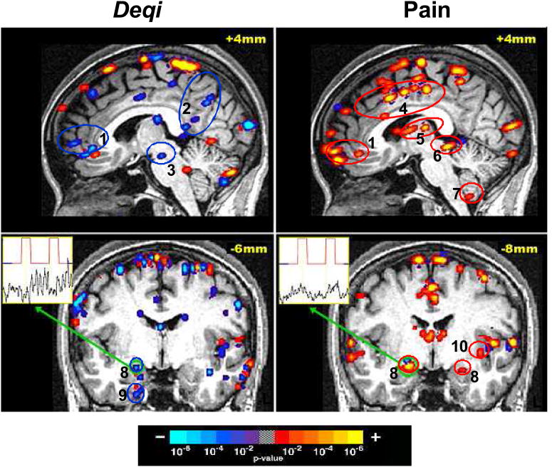
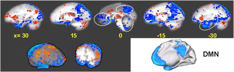
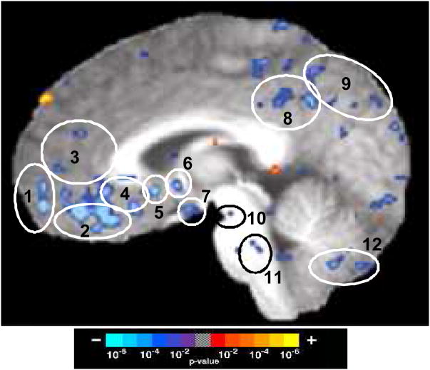
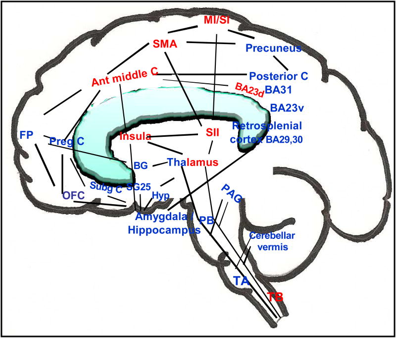
The study of the mechanism of acupuncture action was revolutionized by the use of functional magnetic resonance imaging (fMRI). Over the past decade, our fMRI studies of healthy subjects have contributed substantially to elucidating the central effect of acupuncture on the human brain. These studies have shown that acupuncture stimulation, when associated with sensations comprising deqi, evokes deactivation of a limbic-paralimbic-neocortical network, which encompasses the limbic system, as well as activation of somatosensory brain regions. These networks closely match the default mode network and the anti-correlated task-positive network described in the literature. We have also shown that the effect of acupuncture on the brain is integrated at multiple levels, down to the brainstem and cerebellum. Our studies support the hypothesis that the effect of acupuncture on the brain goes beyond the effect of attention on the default mode network or the somatosensory stimulation of acupuncture needling. The amygdala and hypothalamus, in particular, show decreased activation during acupuncture stimulation that is not commonly associated with default mode network activity. At the same time, our research shows that acupuncture stimulation needs to be done carefully, limiting stimulation when the resulting sensations are very strong or when sharp pain is elicited. When acupuncture induced sharp pain, our studies show that the deactivation was attenuated or reversed in direction. Our results suggest that acupuncture mobilizes the functionally anti-correlated networks of the brain to mediate its actions, and that the effect is dependent on the psychophysical response. In this work we also discuss multiple avenues of future research, including the role of neurotransmitters, the effect of different acupuncture techniques, and the potential clinical application of our research findings to disease states including chronic pain, major depression, schizophrenia, autism, and Alzheimer's disease.


Figures

Figure 1
Figure 1
Time course of acupuncture needling. The acupuncture needle was inserted and the sensitivity of the subject to manipulation was pre-tested and adjusted to tolerance prior to starting functional MRI scanning. The needle remained at rest for 2 min after the start of MRI scanning before bidirectional rotation at 1 Hz for 2 min. The needle was not manipulated for 3 min, then manipulation was repeated for 2 min, followed by a third period of rest for 1min. The needle was removed after MRI scanning was complete. Data analysis compared the blood oxygenation level–dependent (BOLD) MRI signal intensity of the two needling periods with the three rest periods, with the first minute of scanning excluded from analysis to allow for MRI signal equilibration.
Figure 2
Figure 2
The influence of sensations on brain fMRI signal changes during acupuncture and sensory control at ST36. Group average functional results showing signal decreases (blue) and increases (red), thresholded at p<0.001 and a 3-voxel cluster size. All slices are 2mm parasagittal in the right hemisphere in Talairach space (Talairach and Tournoux, 1988). (Left) Acupuncture with deqi sensations without sharp pain (n=11) resulted in widespread signal decreases. (Center) Acupuncture with mixed deqi and sharp pain sensations (n=4) resulted primarily in signal increases. (Right) Sensory control (n=5) also resulted in signal increases beyond dedicated sensorimotor areas. Regions: (1) frontal pole, (2) subgenual cingulate, (3) ventromedial prefrontal cortex, (4) hypothalamus, (5) posterior cingulate, (6) reticular formation, (7) cerebellar vermis, (8) middle cingulate, and (9) thalamus.
Figure 3
Figure 3
Brain activity with acupuncture at ST36 in a single subject who reported deqi sensations without sharp pain during one experimental run and only sharp pain without additional sensations during a subsequent experimental run. The broad degree of deactivation (blue) during acupuncture with deqi is in contrast to the general activation (red) noted during acupuncture with sharp pain alone. Time-course for the voxel with peak signal change within the right amygdala is shown for each run. Regions: (1) pregenual cingulate / frontal pole, (2) posterior cingulate Brodmann area 31 / precuneus, (3) substantia nigra, (4) middle cingulate, (5) thalamus, (6) periaqueductal gray, (7) cerebellar tonsil, (8) amygdala, (9) parahippocampus, (10) insula.
Figure 4
Figure 4
Seed-based cross-correlation analysis of deactivation network in 48 matched subjects in 201 acupuncture runs with deqi and 74 tactile stimulation runs with and without deqi. Reference regions are circled in red, and regions with positive correlation activity are circled in green. Correlations with p<0.001 are shown. (A, B) Correlations for the posterior cingulate Brodmann area 31: (A) Acupuncture: reference voxel (2, −53, 36), showing correlations in the medial prefrontal, posterior medial parietal, medial temporal lobe and temporal pole. (B) Tactile stimulation: reference voxel (3, −63, 31), showing correlations that partially overlap those in acupuncture, but correlations are markedly weaker. (C,D) Correlations for the hypothalamus: (C) Acupuncture: reference voxel (3, −1, −10), showing correlations with regions similar to (A), but more limited in extent. (D) Tactile stimulation: reference voxel (3, −1, −10), showing no regions with significant signal change. Numbered areas: (1) frontal pole and pregenual cingulate, (2) subgenual cingulate, and subgenual area 25 in the ventral route of the medial prefrontal cortex, (3) precuneus, posterior cingulate and retrosplenial cortex of the posterior medial parietal cortex, (4) amygdala, hippocampus, and parahippocampus of the medial temporal lobe, (5) temporal pole, (6) precuneus and posterior cingulate cortex Brodmann area 31, and (7) orbitofrontal cortex, subgenual cingulate, subgenual area SG25 and ventromedial prefrontal cortex.
Figure 5
Figure 5
Comparison of the limbic-paralimbic-neocortical network (LPNN) during acupuncture deqi and the default mode network (DMN) as described in the literature (adapted from Shulman et al., 1997). Multiple sagittal slices through an averaged brain in Talairach space (Talairach and Tournoux, 1988) are shown for the LPNN, spanning both the right hemisphere (left) and the left hemisphere (right), with distance from midline shown in mm. Two brain surface renderings (Cox, 1996) are also shown in the lower left. An oblique view of the DMN is shown in the lower right.
Figure 6
Figure 6
Limbic-Paralimbic-Neocortical Network activity during acupuncture deqi. The averaged BOLD signal changes are shown for 11 subjects during acupuncture at point ST36, showing activation (red) and deactivation (blue). A parasagittal image is shown at 2mm from midline within the right hemisphere in Talairach space (Talairach and Tournoux, 1988). Regions: (1) frontal pole, (2) ventromedial prefrontal cortex, (3) pregenual cingulate, (4) subgenual cingulate, (5) subgenual Brodmann area 25, (6) septal nuclei, (7) hypothalamus, (8) posterior cingulate, 9 precuneus, (10) substantia nigra, (11) reticular formation, and (12) cerebellar vermis.
Figure 7
Figure 7
Functionally anti-correlated task-positive (activation, red) and negative (deactivation, blue) networks involved in acupuncture action shown on a brain sketch, with an overlapping of medial and lateral regions. Regions of deactivation in the limbic–paralimbic–neocortical network aggregate in the medial frontal, parietal and temporal lobes. The basal ganglia, the cerebellar vermis, tonsil and brainstem also show regional deactivation. The cingulate and thalamus have regions in both networks, while the sensorimotor areas, insula, middle cingulate and the dorsal division of posterior cingulate Brodmann area 23 are in the task-positive network. The figure is adapted from Hui et al. (2009). Abbreviations: ant middle C, anterior middle cingulate; BA Brodmann area; BA23d, Brodmann area 23 dorsal; BA23v, Brodmann area 23 ventral; BG, basal ganglia; FP, frontal pole; Hyp, hypothalamus; M1/S1, primary motor/primary sensory cortex; OFC, orbitofrontal cortex; PAG, periaqueductal gray; PB, parabrachial nucleus; preg C, pregenual cingulate; SII, secondary somatosensory cortex; SMA, supplementary motor area; SG25, subgenual area 25; Subg C, subgenual cingulate, TA, tract A; TB, tract B.

References

    1. Anand A, Li Y, Wang Y, Gardner K, Lowe MJ. Reciprocal effects of antidepressant treatment on activity and connectivity of the mood regulating circuit: an FMRI study. J Neuropsychiatry Clin Neurosci. 2007;19:274–282. - PMC - PubMed
    1. Bai L, Qin W, Tian J, Dong M, Pan X, Chen P, Dai J, Yang W, Liu Y. Acupuncture modulates spontaneous activities in the anti-correlated resting brain networks. Brain Res. 2009;1279:37–49. - PubMed
    1. Baliki MN, Geha PY, Apkarian AV, Chialvo DR. Beyond feeling: chronic pain hurts the brain, disrupting the default-mode network dynamics. J Neurosci. 2008;28:1398–1403. - PMC - PubMed
    1. Becerra LR, Breiter HC, Stojanovic M, Fishman S, Edwards A, Comite A, Gonzalez RG, Borsook D. Human brain activation under controlled thermal stimulation and habituation to noxious heat: an fMRI study. Magn Reson Med. 1999;41:1044–1057. - PubMed
    1. Benes FM. Neural circuitry models of schizophrenia: is it dopamine, GABA, glutamate, or something else? Biol Psychiatry. 2009;65:1003–1005. - PubMed

Publication types